Explore
 Validate
 Learn
LearnBMS1043
antibody from Invitrogen Antibodies
Targeting: LGALS3
GALIG, LGALS2, MAC-2
 Western blot
 ELISA Immunocytochemistry Immunoprecipitation Immunohistochemistry Flow cytometry Other assay
Antibody data
- Antibody Data
- Antigen structure
- References [1]
- Comments [0]
- Validations
- Western blot [3]
- Immunocytochemistry [2]
- Other assay [1]
Submit
Validation data
Reference
Comment
Report error
- Product number
- BMS1043 - Provider product page

- Provider
- Invitrogen Antibodies
- Product name
- Galectin 3 Monoclonal Antibody (eBioM3/38 (M3/38)), eBioscience™
- Antibody type
- Monoclonal
- Antigen
- Other
- Description
- Description: The Anti-human Galectin-3 (M3/38) antibody recognizes an epitope in the N-terminal half of lectin. Galectins are a family of animal lectins which appear to exhibit a variety of biological functions. Galectin 3 (aka LGALS3, galactose-specific soluble lectin 3, Mac-2, L-29) is one of the more extensively studied members of this family and is a 30 kDa beta-galactoside-binding protein. Due to a C-terminal carbohydrate binding site, Galectin 3 is capable of binding IgE and mammalian cell surfaces only when homodimerized or homo-oligomerized. Galectin 3 is normally distributed in epithelia of many organs and various inflammatory cells, including macrophages, as well as dendritic cells and Kupffer cells. The expression of this lectin is up- regulated during inflammation, cell proliferation, cell differentiation and through trans-activation by viral proteins. Applications Tested: ELISA, Flow Cytometry, Immunohistochemistry, Immunoprecipitation, Western Blotting.
- Reactivity
- Human
- Host
- Rat
- Isotype
- IgG
- Antibody clone number
- eBioM3/38 (M3/38)
- Vial size
- 100 µg
- Concentration
- 1 mg/mL
- Storage
- -20°C, Avoid Freeze/Thaw Cycles
Submitted references Complement C4 Prevents Viral Infection through Capsid Inactivation.
Bottermann M, Foss S, Caddy SL, Clift D, van Tienen LM, Vaysburd M, Cruickshank J, O'Connell K, Clark J, Mayes K, Higginson K, Lode HE, McAdam MB, Sandlie I, Andersen JT, James LC
Cell host & microbe 2019 Apr 10;25(4):617-629.e7
Cell host & microbe 2019 Apr 10;25(4):617-629.e7
No comments: Submit comment
Supportive validation
- Submitted by
- Invitrogen Antibodies (provider)
- Main image
- Experimental details
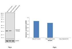
- Knockout of Galectin3 was achieved by CRISPR-Cas9 genome editing using LentiArray™ Lentiviral sgRNA (Product # A32042, Assay ID CRISPR855199_LV and CRISPR855204_LV) and LentiArray Cas9 Lentivirus (Product # A32064). Western blot analysis of Galectin3 was performed by loading 30 µg of HeLa Wild Type (Lane 1), HeLa Cas9 (Lane 2) andHela Galectin3 KO (Lane 3) membrane enriched extracts. The samples were electrophoresed using NuPAGE™ Novex™ 4-12% Bis-Tris Protein Gel (Product # NP0322BOX). Resolved proteins were then transferred onto a nitrocellulose membrane (Product # IB23001) by iBlot® 2 Dry Blotting System (Product # IB21001). The blot was probed with Anti-Galectin 3 Monoclonal Antibody (eBioM3/38 (M3/38)), eBioscience™ (Product # BMS1043, 0.25 µg/mL dilution) and F(ab)2-Rabbit anti-Rat IgG (H+L) Secondary Antibody, HRP (Product # PA1-29927, 1:6,000 dilution) using the iBright FL 1000 (Product # A32752). Chemiluminescent detection was performed using Novex® ECL Chemiluminescent Substrate Reagent Kit (Product # WP20005). Loss of signal upon CRISPR mediated knockout (KO) using the LentiArray™ CRISPR product line confirms that antibody is specific to Galectin3.

- Submitted by
- Invitrogen Antibodies (provider)
- Main image
- Experimental details
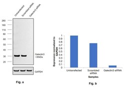
- Knockdown of Galectin 3 was achieved by transfecting MDA-MB-231 cells with Galectin (LGALS3) specific siRNAs (Silencer® select Product # S8149, S8148). Western blot analysis of Galectin 3 Monoclonal Antibody (eBioM3/38 (M3/38)) (Product # BMS1043) (Fig. a) was performed using whole cell extracts from the knockdown cells (Lane 3), non-specific scrambled siRNA transfected cells (Lane 2) and untransfected cells (Lane 1). The blot was probed with the primary antibody (1 µg/mL) and detected by chemiluminescence with F(ab)2-Rabbit anti-Rat IgG (H+L) Secondary Antibody, HRP, Superclonal™ Recombinant Secondary Antibody, HRP (Product # PA1-29927, 0.25 µg/mL, 1:4000 dilution) using the iBright FL 1000 (Product # A32752). Densitometric analysis of this western blot is shown in histogram (Fig. b). Decrease in signal upon siRNA mediated knock down confirms that antibody is specific to Galectin 3.

- Submitted by
- Invitrogen Antibodies (provider)
- Main image
- Experimental details
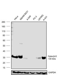
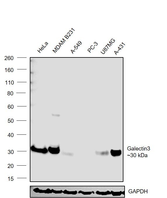
- Western blot was performed using Anti-Galectin 3 Monoclonal antibody (Product # BMS1043) and a band around 30 kDa corresponding to Galectin 3 was observed in cell lines tested. Whole cell extracts (30 µg lysate) of HeLa (Lane 1), MDA-MB-231 (Lane 2), A-549 (Lane 3), PC-3 (Lane 4), U-87 mg (Lane 5) and A-431 (Lane 6) were electrophoresed using NuPAGE® 4-12 % Bis-Tris gel (Product # NP0322BOX). Resolved proteins were then transferred onto a nitrocellulose membrane (Product # IB23001) by iBlot® 2 Dry Blotting System (Product # IB21001). The blot was probed with the primary antibody (1 µg/mL) and detected by chemiluminescence with F(ab)2-Rabbit anti-Rat IgG (H+L) Secondary Antibody, HRP, Superclonal™ Recombinant Secondary Antibody, HRP (Product # PA1-29927), 0.25 µg/mL, 1:4000 dilution) using the iBright FL 1000 (Product # A32752). Chemiluminescent detection was performed using Novex® ECL Chemiluminescent Substrate Reagent Kit (Product # WP20005).
Supportive validation
- Submitted by
- Invitrogen Antibodies (provider)
- Main image

- Experimental details
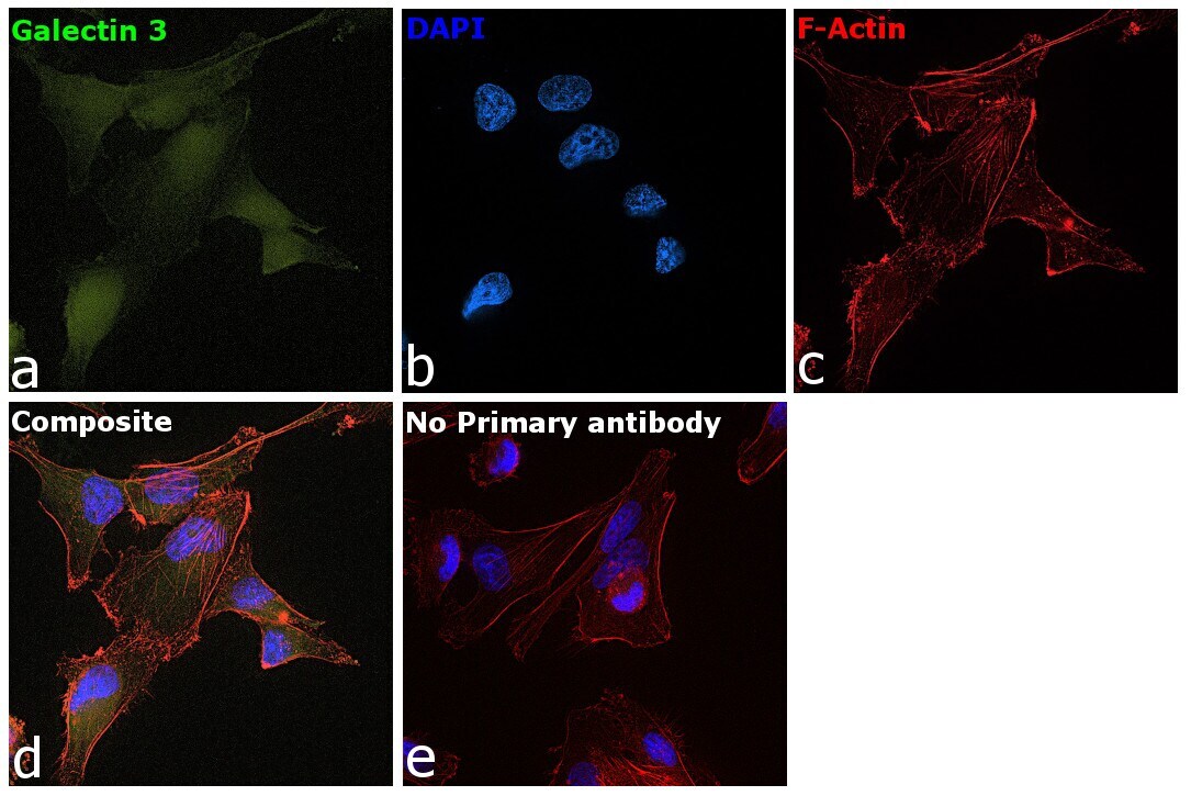
- Immunofluorescence analysis of Galectin3 was performed using 70% confluent log phase MDA-MB-231 cells. The cells were fixed with 4% Paraformaldehyde for 10 minutes, permeabilized with 0.1% Triton™ X-100 for 10 minutes, and blocked with 2% BSA for 45 minutes at room temperature. The cells were labeled with Galectin 3 Monoclonal Antibody (eBioM3/38 (M3/38)) (Product # BMS1043) at 5 µg/mL in 0.1% BSA, incubated at 4 degree celsius overnight and then labeled with Goat anti-Rat IgG (H+L) Cross-Adsorbed Secondary Antibody, Alexa Fluor 488 (Product # A-11006 , 1:2000 dilution) for 45 minutes at room temperature (Panel a: Green). Nuclei (Panel b: Blue) were stained with ProLong™ Diamond Antifade Mountant with DAPI (Product # P36962). F-actin (Panel c: Red) was stained with Rhodamine Phalloidin (Product # R415, 1:300 dilution). Panel d represents the merged image showing peri-nuclear and nuclear localization. Panel e represents control cells with no primary antibody to assess background. The images were captured at 60X magnification.

- Submitted by
- Invitrogen Antibodies (provider)
- Main image

- Experimental details
- Immunofluorescence analysis of Galectin3 was performed using 70% confluent log phase MDA-MB-231 cells. The cells were fixed with 4% Paraformaldehyde for 10 minutes, permeabilized with 0.1% Triton™ X-100 for 10 minutes, and blocked with 2% BSA for 45 minutes at room temperature. The cells were labeled with Galectin 3 Monoclonal Antibody (eBioM3/38 (M3/38)) (Product # BMS1043) at 5 µg/mL in 0.1% BSA, incubated at 4 degree celsius overnight and then labeled with Goat anti-Rat IgG (H+L) Cross-Adsorbed Secondary Antibody, Alexa Fluor 488 (Product # A-11006 , 1:2000 dilution) for 45 minutes at room temperature (Panel a: Green). Nuclei (Panel b: Blue) were stained with ProLong™ Diamond Antifade Mountant with DAPI (Product # P36962). F-actin (Panel c: Red) was stained with Rhodamine Phalloidin (Product # R415, 1:300 dilution). Panel d represents the merged image showing peri-nuclear and nuclear localization. Panel e represents control cells with no primary antibody to assess background. The images were captured at 60X magnification.
Supportive validation
- Submitted by
- Invitrogen Antibodies (provider)
- Main image
- Experimental details
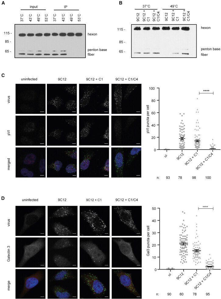
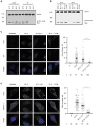
- Figure 4 C1 and C4 Prevent Adenoviral Protein VI Exposure and Endosomal Escape (A) IP of Ad5 with 9C12 after Ad5 was incubated at the indicated temperatures for 30 min. WB: anti-adenovirus. (B) IP after Ad5 and 9C12 were complexed with C1 or C1/C4 and then incubated at 37degC or 49degC for 30 min. WB: anti-adenovirus. (C and D) HeLa TRIM21 KO cells were infected for 30 min in the presence of 9C12 or 9C12+ complement. Error bars depict the mean +- SEM of the indicated number of cells (n) acquired in three independent experiments. Scale bar, 5 mum. (C) Left: Ad5 staining is displayed in green; protein VI staining is depicted in red. Right: quantification of protein VI puncta per cell in the indicated conditions. (D) Left: Ad5 staining is displayed in green; Galectin-3 staining is depicted in red. Right: quantification of Galectin-3 puncta per cell in the indicated conditions. Original western blots are included in Figure S6 .