Explore
 Validate
 Learn
Learn Western blot
Antibody data
- Antibody Data
- Antigen structure
- References [2]
- Comments [0]
- Validations
- Western blot [2]
- Immunohistochemistry [1]
Submit
Validation data
Reference
Comment
Report error
- Product number
- AF6895 - Provider product page

- Provider
- R&D Systems
- Product name
- Mouse/Rat Nesfatin-1/Nucleobindin-2 Antibody
- Antibody type
- Polyclonal
- Description
- Immunogen affinity purified. Detects rat Nesfatin-1/Nucleobindin-2 in direct ELISAs and mouse and rat Nesfatin-1/Nucleobindin-2 in Western blots. In direct ELISAs, approximately 7% cross-reactivity with recombinant human Nesfatin-1/Nucleobindin-2 is observed.
- Reactivity
- Mouse, Rat
- Host
- Sheep
- Conjugate
- Unconjugated
- Antigen sequence
Q9JI85- Isotype
- IgG
- Vial size
- 100 ug
- Concentration
- LYOPH
- Storage
- Use a manual defrost freezer and avoid repeated freeze-thaw cycles. 12 months from date of receipt, -20 to -70 °C as supplied. 1 month, 2 to 8 °C under sterile conditions after reconstitution. 6 months, -20 to -70 °C under sterile conditions after reconstitution.
Submitted references Loss of Nucleobindin-2 Causes Insulin Resistance in Obesity without Impacting Satiety or Adiposity.
Role of Nesfatin-1 in the Reproductive Axis of Male Rat.
Ravussin A, Youm YH, Sander J, Ryu S, Nguyen K, Varela L, Shulman GI, Sidorov S, Horvath TL, Schultze JL, Dixit VD
Cell reports 2018 Jul 31;24(5):1085-1092.e6
Cell reports 2018 Jul 31;24(5):1085-1092.e6
Role of Nesfatin-1 in the Reproductive Axis of Male Rat.
Gao X, Zhang K, Song M, Li X, Luo L, Tian Y, Zhang Y, Li Y, Zhang X, Ling Y, Fang F, Liu Y
Scientific reports 2016 Sep 7;6:32877
Scientific reports 2016 Sep 7;6:32877
No comments: Submit comment
Supportive validation
- Submitted by
- R&D Systems (provider)
- Main image
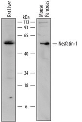
- Experimental details
- Detection of Mouse and Rat Nesfatin-1/Nucleobindin-2 by Western Blot. Western blot shows lysates of rat liver tissue and mouse pancreas tissue. PVDF membrane was probed with 2 µg/mL of Sheep Anti-Mouse/Rat Nesfatin-1/Nucleobindin-2 Antigen Affinity-purified Polyclonal Antibody (Catalog # AF6895) followed by HRP-conjugated Anti-Sheep IgG Secondary Antibody (Catalog # HAF016). A specific band was detected for Nesfatin-1/Nucleobindin-2 at approximately 50 to 55 kDa (as indicated). This experiment was conducted under reducing conditions and using Immunoblot Buffer Group 1.

- Submitted by
- R&D Systems (provider)
- Main image
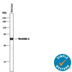
- Experimental details
- Detection of Rat Nesfatin-1/Nucleobindin-2 by Simple WesternTM. Simple Western lane view shows lysates of rat liver tissue, loaded at 0.2 mg/mL. A specific band was detected for Nesfatin-1/ Nucleobindin-2 at approximately 55 kDa (as indicated) using 20 µg/mL of Sheep Anti-Mouse/Rat Nesfatin-1/Nucleobindin-2 Antigen Affinity-purified Polyclonal Antibody (Catalog # AF6895) followed by 1:50 dilution of HRP-conjugated Anti-Sheep IgG Secondary Antibody (Catalog # HAF016). This experiment was conducted under reducing conditions and using the 12-230 kDa separation system.

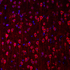
Supportive validation
- Submitted by
- R&D Systems (provider)
- Main image
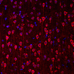
- Experimental details
- Nesfatin-1/Nucleobindin-2 in Rat Brain. Nesfatin-1/Nucleobindin-2 was detected in immersion fixed frozen sections of rat brain (cortex) using Sheep Anti-Mouse/Rat Nesfatin-1/Nucleobindin-2 Antigen Affinity-purified Polyclonal Antibody (Catalog # AF6895) at 1.7 µg/mL overnight at 4 °C. Tissue was stained using the NorthernLights™ 557-conjugated Anti-Sheep IgG Secondary Antibody (red; Catalog # NL010) and counterstained with DAPI (blue). Specific staining was localized to neuronal cell bodies and processes. View our protocol for Fluorescent IHC Staining of Frozen Tissue Sections.