Explore
 Validate
 Learn
Learn Western blot
 ELISA
ELISAAntibody data
- Antibody Data
- Antigen structure
- References [0]
- Comments [0]
- Validations
- Western blot [2]
- Immunocytochemistry [1]
- Flow cytometry [1]
Submit
Validation data
Reference
Comment
Report error
- Product number
- 600-401-GU3 - Provider product page

- Provider
- Invitrogen Antibodies
- Product name
- xCT Polyclonal Antibody
- Antibody type
- Polyclonal
- Antigen
- Synthetic peptide
- Reactivity
- Human
- Host
- Rabbit
- Isotype
- IgG
- Vial size
- 100 µg
- Concentration
- 1.02 mg/mL
- Storage
- -20° C, Avoid Freeze/Thaw Cycles
No comments: Submit comment
Supportive validation
- Submitted by
- Invitrogen Antibodies (provider)
- Main image
- Experimental details
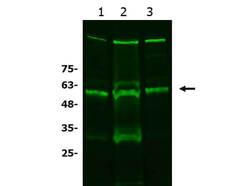
- Western Blot of Rabbit anti-xCT antibody. Lane 1: A549 WCL (p/n W09-001-372). Lane 2: HCT-116 WCL (p/n W09-001-GM4). Lane 3: HeLa WCL (p/n W09-000-364). Load: 10 µg per lane. Primary antibody: xCT antibody at 1:1000 for overnight at 4°C. Secondary antibody: donkey anti-rabbit secondary DyLight™488 antibody (p/n 611-741-127) at 1:20,000 for one hour at RT. Block: BlockOut blocking buffer (p/n MB-073) one hour at RT. Predicted/Observed size: 56 kDa. Other band(s): xCT processing caused by dimerization, glycosylation, and/or phosphorylation.
- Submitted by
- Invitrogen Antibodies (provider)
- Main image
- Experimental details
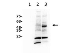
- Western Blot of Rabbit anti-xCT antibody. Lane 1: recombinant BCL3 (unrelated negative control). Lane 2: NIH 3T3 WCL (p/n W10-000-358). Lane 3: A549 WCL (p/n W09-001-372). Load: 10 µg per lane. Primary antibody: xCT antibody at 1:1000 for overnight at 4°C. Secondary antibody: goat anti-rabbit secondary HRP antibody (p/n 611-103-122) at 1:20,000 for one hour at RT. Block: 5% BSA blocking buffer one hour at RT. Predicted/Observed size: 56 kDa. Other band(s): xCT processing caused by dimerization, glycosylation, and/or phosphorylation.

Supportive validation
- Submitted by
- Invitrogen Antibodies (provider)
- Main image
- Experimental details
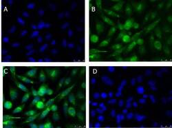
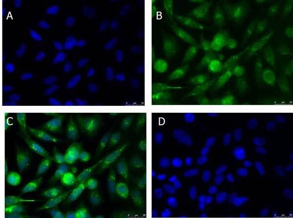
- Immunofluorescence Microscopy of Rabbit anti-xCT antibody. Tissue: PC3 cells. Fixation: 100% MeOH. Antigen retrieval: not required. Primary antibody: xCT antibody at 10 µg/mL overnight at 4°C. Secondary antibody: Donkey Anti-Rabbit IgG DyLight™ 488 (p/n 611-741-127) at 5 µg/mL for 2 h at RT. Localization: xCT is localized on the cell membrane and vesicles. Staining: xCT as green fluorescent signal with DAPI (blue) nuclear counterstain.
Supportive validation
- Submitted by
- Invitrogen Antibodies (provider)
- Main image
- Experimental details
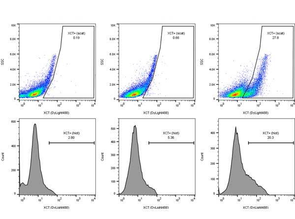
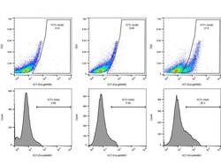
- Flow Cytometry of rabbit anti-xCT antibody. Cells: breast carcinoma cells Primary antibody: xCT antibody at 1.0 µg/mL for one hour at 4°C. Secondary antibody: Donkey anti-Rabbit IgG Dylight™488 Antibody p/n (611-741-127) at 1 µg/ml in 200 ul for one hour on ice.