Explore
 Validate
 Learn
Learn Western blot
 Immunocytochemistry
Antibody data
- Antibody Data
- Antigen structure
- References [2]
- Comments [0]
- Validations
- Immunocytochemistry [5]
- Immunohistochemistry [1]
- Other assay [2]
Submit
Validation data
Reference
Comment
Report error
- Product number
- PA5-32228 - Provider product page

- Provider
- Invitrogen Antibodies
- Product name
- SHMT2 Polyclonal Antibody
- Antibody type
- Polyclonal
- Antigen
- Recombinant full-length protein
- Description
- Recommended positive controls: 293T, HeLa, A431, HepG2. Predicted reactivity: Mouse (93%), Rat (91%), Xenopus laevis (81%), Rabbit (93%), Bovine (92%). Store product as a concentrated solution. Centrifuge briefly prior to opening the vial.
- Reactivity
- Human, Mouse, Rat
- Host
- Rabbit
- Isotype
- IgG
- Vial size
- 100 μL
- Concentration
- 1 mg/mL
- Storage
- Store at 4°C short term. For long term storage, store at -20°C, avoiding freeze/thaw cycles.
Submitted references Shmt2: A Stat3 Signaling New Player in Prostate Cancer Energy Metabolism.
Mitochondrial dysfunction remodels one-carbon metabolism in human cells.
Marrocco I, Altieri F, Rubini E, Paglia G, Chichiarelli S, Giamogante F, Macone A, Perugia G, Magliocca FM, Gurtner A, Maras B, Ragno R, Patsilinakos A, Manganaro R, Eufemi M
Cells 2019 Sep 6;8(9)
Cells 2019 Sep 6;8(9)
Mitochondrial dysfunction remodels one-carbon metabolism in human cells.
Bao XR, Ong SE, Goldberger O, Peng J, Sharma R, Thompson DA, Vafai SB, Cox AG, Marutani E, Ichinose F, Goessling W, Regev A, Carr SA, Clish CB, Mootha VK
eLife 2016 Jun 16;5
eLife 2016 Jun 16;5
No comments: Submit comment
Supportive validation
- Submitted by
- Invitrogen Antibodies (provider)
- Main image

- Experimental details
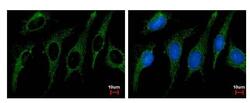
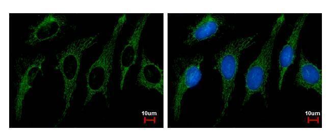
- SHMT2 Polyclonal Antibody detects SHMT2 protein at Mitochondria by immunofluorescent analysis. Sample: HeLa cells were fixed in ice-cold MeOH for 1 sec. Green: SHMT2 protein stained by SHMT2 Polyclonal Antibody (Product # PA5-32228) diluted at 1:500. Blue: Hoechst 33342 staining.

- Submitted by
- Invitrogen Antibodies (provider)
- Main image

- Experimental details
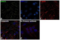
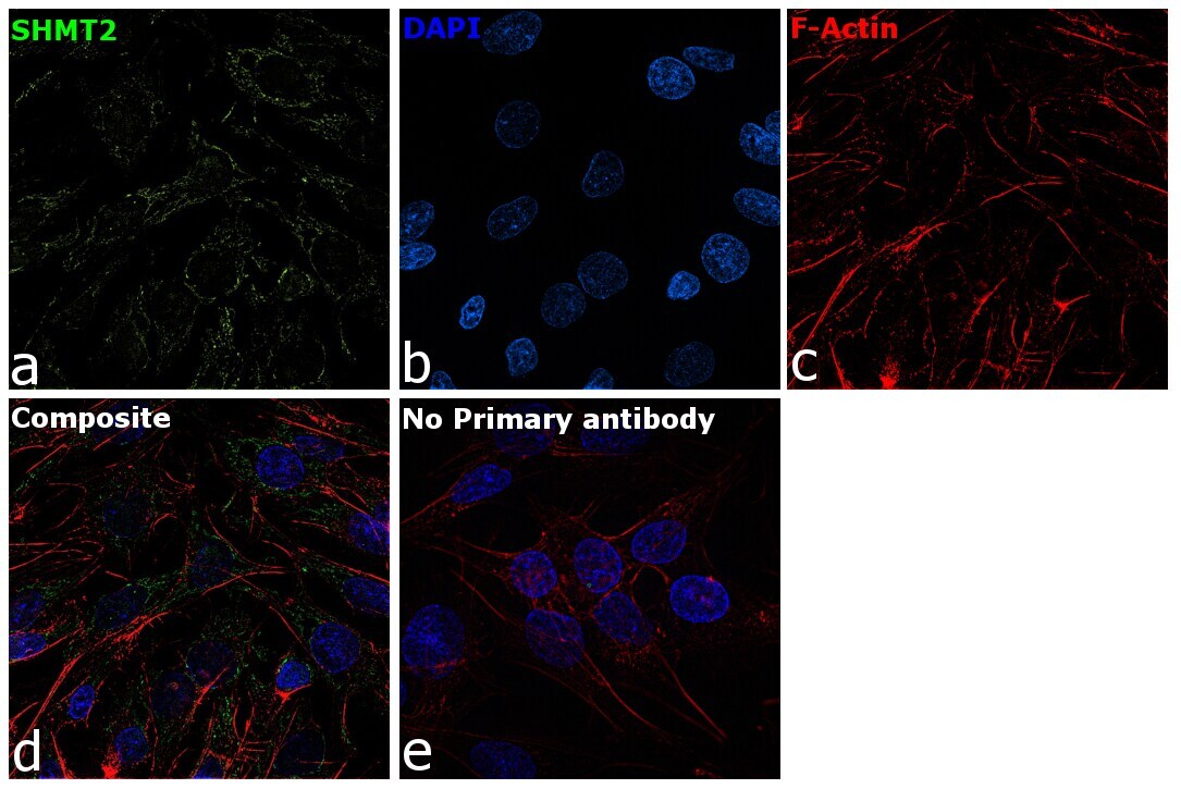
- Immunofluorescence analysis of Serine hydroxymethyltransferase, mitochondrial was performed using 70% confluent log phase Hep G2 cells. The cells were fixed with 4% paraformaldehyde for 10 minutes, permeabilized with 0.1% Triton™ X-100 for 15 minutes, and blocked with 2% BSA for 45 minutes at room temperature. The cells were labeled with SHMT2 Polyclonal Antibody (Product # PA5-32228) at 1:100 dilution in 0.1% BSA, incubated at 4 degree celsius overnight and then labeled with Goat anti-Rabbit IgG (H+L) Superclonal™ Recombinant Secondary Antibody, Alexa Fluor® 488 conjugate (Product # A27034), (1:2000 dilution), for 45 minutes at room temperature (Panel a: Green). Nuclei (Panel b:Blue) were stained with ProLong™ Diamond Antifade Mountant with DAPI (Product # P36962). F-actin (Panel c: Red) was stained with Rhodamine Phalloidin (Product # R415, 1:300). Panel d represents the merged image showing Mitochondrial localization. Panel e represents control cells with no primary antibody to assess background. The images were captured at 60X magnification.

- Submitted by
- Invitrogen Antibodies (provider)
- Main image
- Experimental details
- SHMT2 Polyclonal Antibody detects SHMT2 protein at Mitochondria by immunofluorescent analysis. Sample: HeLa cells were fixed in ice-cold MeOH for 1 sec. Green: SHMT2 protein stained by SHMT2 Polyclonal Antibody (Product # PA5-32228) diluted at 1:500. Blue: Hoechst 33342 staining.
- Submitted by
- Invitrogen Antibodies (provider)
- Main image
- Experimental details
- Immunofluorescence analysis of Serine hydroxymethyltransferase, mitochondrial was performed using 70% confluent log phase Hep G2 cells. The cells were fixed with 4% paraformaldehyde for 10 minutes, permeabilized with 0.1% Triton™ X-100 for 15 minutes, and blocked with 2% BSA for 45 minutes at room temperature. The cells were labeled with SHMT2 Polyclonal Antibody (Product # PA5-32228) at 1:100 dilution in 0.1% BSA, incubated at 4 degree celsius overnight and then labeled with Goat anti-Rabbit IgG (Heavy Chain) Superclonal™ Recombinant Secondary Antibody, Alexa Fluor® 488 conjugate (Product # A27034), (1:2000 dilution), for 45 minutes at room temperature (Panel a: Green). Nuclei (Panel b:Blue) were stained with ProLong™ Diamond Antifade Mountant with DAPI (Product # P36962). F-actin (Panel c: Red) was stained with Rhodamine Phalloidin (Product # R415, 1:300). Panel d represents the merged image showing Mitochondrial localization. Panel e represents control cells with no primary antibody to assess background. The images were captured at 60X magnification.
Supportive validation
- Submitted by
- Invitrogen Antibodies (provider)
- Main image
- Experimental details
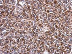
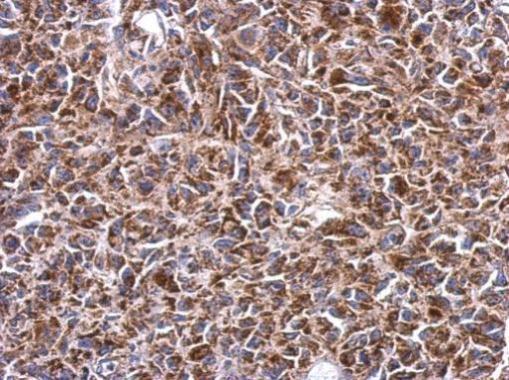
- Immunohistochemical analysis of paraffin-embedded HeLa xenograft, using SHMT2 (Product # PA5-32228) antibody at 1:500 dilution. Antigen Retrieval: EDTA based buffer, pH 8.0, 15 min.
Supportive validation
- Submitted by
- Invitrogen Antibodies (provider)
- Main image
- Experimental details
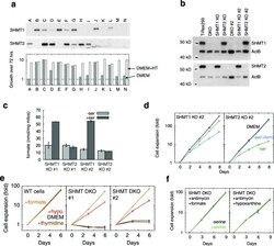
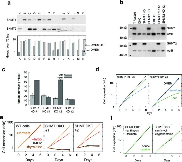
- Figure 5--figure supplement 2. SHMT1 and SHMT2 single and double knockouts. ( a ) Western blots (top) and growth rates (bottom) for fourteen single cell clones derived from simultaneous CRISPR transfection for SHMT1 and SHMT2 knockout. All clones were expanded in the presence of hypoxanthine and thymidine (HT). ( b ) Western blots showing SHMT1 and SHMT2 single and double knockouts. ( c ) Formate synthesis from mitochondria isolated from SHMT1 and SHMT2 knockout cell lines. ( d ) Replicate serine dependence data from single knockouts. ( e ) Growth of SHMT1/SHMT2 double KO cells (two independent clones) with different 1C-related supplements. ( f ) 1C-rescued double knockouts do not show serine dependence, even when treated with RC inhibitor. n = 1 for panel a; n = 3 for panels c-f . DOI: http://dx.doi.org/
- Submitted by
- Invitrogen Antibodies (provider)
- Main image
- Experimental details
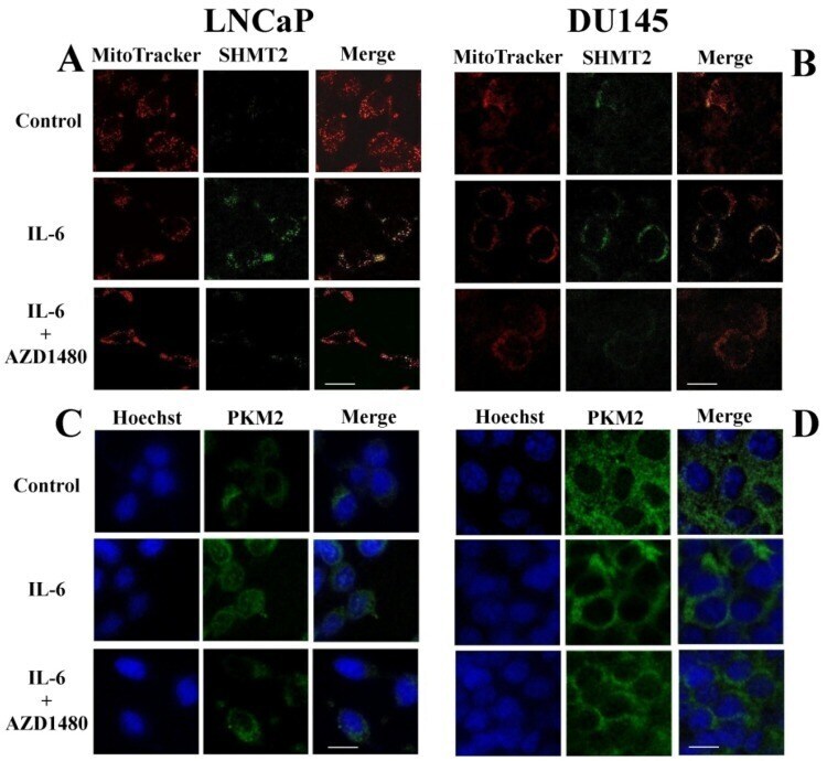
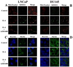
- Figure 7 Cellular redistribution of SHMT2 and PKM2 following IL-6 stimulation. Immunofluorescence analysis of SHMT2 ( A , B ) and PKM2 ( C , D ) proteins in LNCaP and DU145 cells after 4 h of IL-6 stimulation, with or without a Jak2 inhibitor (AZD1480). ( A , C ) LNCaP cell line. ( B , D ) DU145 cell line. The images were captured with a confocal fluorescence microscope (ZEISS LSM510META) using 60x oil immersion objective (scale bar 10 mum).