Explore
 Validate
 Learn
Learn Immunohistochemistry
Antibody data
- Antibody Data
- Antigen structure
- References [2]
- Comments [0]
- Validations
- Immunohistochemistry [1]
- Other assay [1]
Submit
Validation data
Reference
Comment
Report error
- Product number
- PA5-61896 - Provider product page

- Provider
- Invitrogen Antibodies
- Product name
- RELM beta Polyclonal Antibody
- Antibody type
- Polyclonal
- Antigen
- Recombinant protein fragment
- Description
- Immunogen sequence: LDSVMDKKIK DVLNSLEYSP SPISKKLSCA SVKSQGRPSS CPAGMAVTGC ACGYGCGSWD VQLETTCHCQ CSVVDWTTAR CCHLT Highest antigen sequence identity to the following orthologs: Mouse - 56%, Rat - 60%.
- Reactivity
- Human
- Host
- Rabbit
- Isotype
- IgG
- Vial size
- 100 μL
- Concentration
- 0.1 mg/mL
- Storage
- Store at 4°C short term. For long term storage, store at -20°C, avoiding freeze/thaw cycles.
Submitted references Comparison of the host response to larvicidal and nonlarvicidal treatment of naturally acquired cyathostomin infections in horses.
Building a Thick Mucus Hydrogel Layer to Improve the Physiological Relevance of In Vitro Primary Colonic Epithelial Models.
Steuer AE, Scoggin K, Stewart JC, Barker VD, Adams AA, Loynachan AT, Nielsen MK
Parasite immunology 2022 Oct;44(10):e12941
Parasite immunology 2022 Oct;44(10):e12941
Building a Thick Mucus Hydrogel Layer to Improve the Physiological Relevance of In Vitro Primary Colonic Epithelial Models.
Wang Y, Kim R, Sims CE, Allbritton NL
Cellular and molecular gastroenterology and hepatology 2019;8(4):653-655.e5
Cellular and molecular gastroenterology and hepatology 2019;8(4):653-655.e5
No comments: Submit comment
Supportive validation
- Submitted by
- Invitrogen Antibodies (provider)
- Main image
- Experimental details
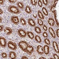
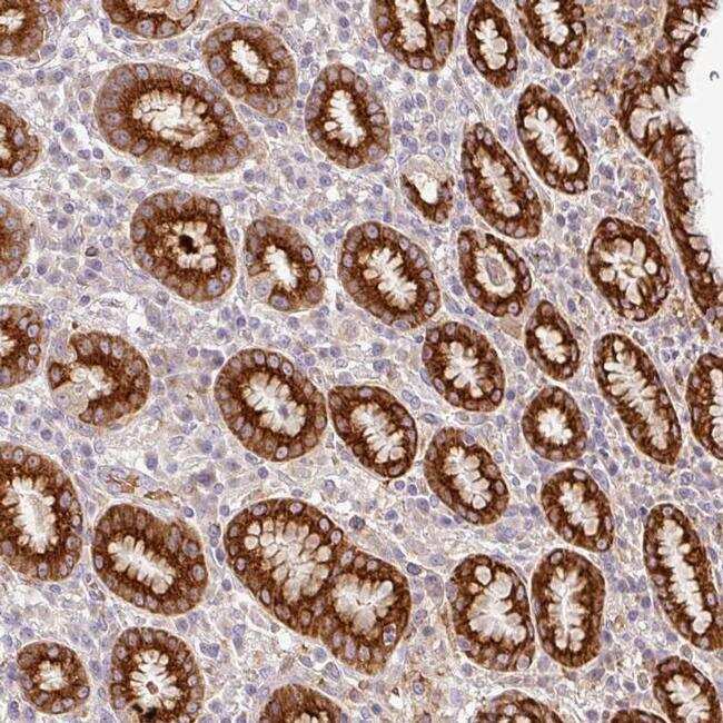
- Immunohistochemical analysis of RELM beta in human stomach using RELM beta Polyclonal Antibody (Product # PA5-61896) shows strong cytoplasmic positivity in glandular cells.
Supportive validation
- Submitted by
- Invitrogen Antibodies (provider)
- Main image
- Experimental details
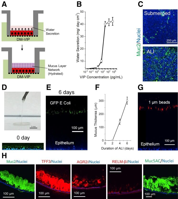
- Figure 1 VIP-assisted ALI culture generates physiologic mucus layer above intestinal epithelial cells. ( A ) Culture schematic. ( B ) Dose-dependent water secretion of VIP at 24 hours. ( C ) Immunofluorescence of monolayers differentiated under submerged and ALI conditions in the presence of VIP. Green: anti-Muc2; blue: Hoechst 33342. ( D ) Removal of hydrated mucus with forceps. ( E ) Side-view confocal micrograph showing tissues with mucus separating bacteria from epithelial cells at 0 and 6 days, respectively. Green: green fluorescent protein (GFP)-expressing Escherichia coli ; blue: Hoechst 33342. ( F ) Plot of mucus thickness vs duration of ALI. ( G ) Representative side-view confocal micrograph showing 1 mum red fluorescent beads separated from epithelial cells by mucus. ( H ) Immunofluorescence staining of paraffin-embedded, sectioned monolayers. DM, differentiation medium.