Explore
 Validate
 Learn
Learn Western blot
 Immunocytochemistry
Antibody data
- Antibody Data
- Antigen structure
- References [2]
- Comments [0]
- Validations
- Immunocytochemistry [1]
Submit
Validation data
Reference
Comment
Report error
- Product number
- AF1187 - Provider product page

- Provider
- R&D Systems
- Product name
- Mouse TREM-1 Antibody
- Antibody type
- Polyclonal
- Description
- Antigen Affinity-purified. Detects mouse TREM-1 in ELISAs and Western blots. In sandwich immunoassays, less than 0.2% cross-reactivity with recombinant human (rh) TREM-1 is observed.
- Reactivity
- Mouse
- Host
- Goat
- Conjugate
- Unconjugated
- Antigen sequence
Q9JKE2- Isotype
- IgG
- Vial size
- 100 ug
- Concentration
- LYOPH
- Storage
- Use a manual defrost freezer and avoid repeated freeze-thaw cycles. 12 months from date of receipt, -20 to -70 °C as supplied. 1 month, 2 to 8 °C under sterile conditions after reconstitution. 6 months, -20 to -70 °C under sterile conditions after reconstitution.
Submitted references TREM-1 amplifies corneal inflammation after Pseudomonas aeruginosa infection by modulating Toll-like receptor signaling and Th1/Th2-type immune responses.
TREM-1--expressing intestinal macrophages crucially amplify chronic inflammation in experimental colitis and inflammatory bowel diseases.
Wu M, Peng A, Sun M, Deng Q, Hazlett LD, Yuan J, Liu X, Gao Q, Feng L, He J, Zhang P, Huang X
Infection and immunity 2011 Jul;79(7):2709-16
Infection and immunity 2011 Jul;79(7):2709-16
TREM-1--expressing intestinal macrophages crucially amplify chronic inflammation in experimental colitis and inflammatory bowel diseases.
Schenk M, Bouchon A, Seibold F, Mueller C
The Journal of clinical investigation 2007 Oct;117(10):3097-106
The Journal of clinical investigation 2007 Oct;117(10):3097-106
No comments: Submit comment
Supportive validation
- Submitted by
- R&D Systems (provider)
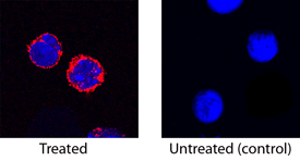
- Main image

- Experimental details
- TREM-1 in Mouse Splenocytes. TREM-1 was detected in immersion fixed mouse splenocytes treated with LPS (left panel) or untreated (right panel) using Goat Anti-Mouse TREM-1 Antigen Affinity-purified Polyclonal Antibody (Catalog # AF1187) at 15 µg/mL for 3 hours at room temperature. Cells were stained using the NorthernLights™ 557-conjugated Anti-Goat IgG Secondary Antibody (red; Catalog # NL001) and counterstained with DAPI (blue). Specific staining was localized to cytoplasm. View our protocol for Fluorescent ICC Staining of Non-adherent Cells.