Explore
 Validate
 Learn
Learn Western blot
 Immunoprecipitation
Antibody data
- Antibody Data
- Antigen structure
- References [8]
- Comments [0]
- Validations
- Western blot [3]
- Immunocytochemistry [2]
- Immunohistochemistry [1]
- Other assay [2]
Submit
Validation data
Reference
Comment
Report error
- Product number
- MA5-13794 - Provider product page

- Provider
- Invitrogen Antibodies
- Product name
- Cdc25A Monoclonal Antibody (DCS-120)
- Antibody type
- Monoclonal
- Antigen
- Purifed from natural sources
- Description
- MA5-13794 targets CDC25A in IP, IHC (P) and WB applications and shows reactivity with Human and Rat samples. The MA5-13794 immunogen is purified recombinant CDC25A.
- Reactivity
- Human, Mouse, Rat
- Host
- Mouse
- Isotype
- IgG
- Antibody clone number
- DCS-120
- Vial size
- 500 µL
- Concentration
- 0.2 mg/mL
- Storage
- 4° C
Submitted references Androgen Receptor, Although Not a Specific Marker For, Is a Novel Target to Suppress Glioma Stem Cells as a Therapeutic Strategy for Glioblastoma.
The miRNA-449 family mediates doxorubicin resistance in triple-negative breast cancer by regulating cell cycle factors.
Targeting radioresistant breast cancer cells by single agent CHK1 inhibitor via enhancing replication stress.
A subset of cancer cell lines is acutely sensitive to the Chk1 inhibitor MK-8776 as monotherapy due to CDK2 activation in S phase.
CDC25A protein stability represents a previously unrecognized target of HER2 signaling in human breast cancer: implication for a potential clinical relevance in trastuzumab treatment.
The let-7 microRNA represses cell proliferation pathways in human cells.
Differential utilization of cyclin D1 and cyclin D3 in the distinct mitogenic stimulations by growth factors and TSH of human thyrocytes in primary culture.
SCFbeta-TRCP links Chk1 signaling to degradation of the Cdc25A protein phosphatase.
Zhao N, Wang F, Ahmed S, Liu K, Zhang C, Cathcart SJ, DiMaio DJ, Punsoni M, Guan B, Zhou P, Wang S, Batra SK, Bronich T, Hei TK, Lin C, Zhang C
Frontiers in oncology 2021;11:616625
Frontiers in oncology 2021;11:616625
The miRNA-449 family mediates doxorubicin resistance in triple-negative breast cancer by regulating cell cycle factors.
Tormo E, Ballester S, Adam-Artigues A, Burgués O, Alonso E, Bermejo B, Menéndez S, Zazo S, Madoz-Gúrpide J, Rovira A, Albanell J, Rojo F, Lluch A, Eroles P
Scientific reports 2019 Mar 29;9(1):5316
Scientific reports 2019 Mar 29;9(1):5316
Targeting radioresistant breast cancer cells by single agent CHK1 inhibitor via enhancing replication stress.
Zhang Y, Lai J, Du Z, Gao J, Yang S, Gorityala S, Xiong X, Deng O, Ma Z, Yan C, Susana G, Xu Y, Zhang J
Oncotarget 2016 Jun 7;7(23):34688-702
Oncotarget 2016 Jun 7;7(23):34688-702
A subset of cancer cell lines is acutely sensitive to the Chk1 inhibitor MK-8776 as monotherapy due to CDK2 activation in S phase.
Sakurikar N, Thompson R, Montano R, Eastman A
Oncotarget 2016 Jan 12;7(2):1380-94
Oncotarget 2016 Jan 12;7(2):1380-94
CDC25A protein stability represents a previously unrecognized target of HER2 signaling in human breast cancer: implication for a potential clinical relevance in trastuzumab treatment.
Brunetto E, Ferrara AM, Rampoldi F, Talarico A, Cin ED, Grassini G, Spagnuolo L, Sassi I, Ferro A, Cuorvo LV, Barbareschi M, Piccinin S, Maestro R, Pecciarini L, Doglioni C, Cangi MG
Neoplasia (New York, N.Y.) 2013 Jun;15(6):579-90
Neoplasia (New York, N.Y.) 2013 Jun;15(6):579-90
The let-7 microRNA represses cell proliferation pathways in human cells.
Johnson CD, Esquela-Kerscher A, Stefani G, Byrom M, Kelnar K, Ovcharenko D, Wilson M, Wang X, Shelton J, Shingara J, Chin L, Brown D, Slack FJ
Cancer research 2007 Aug 15;67(16):7713-22
Cancer research 2007 Aug 15;67(16):7713-22
Differential utilization of cyclin D1 and cyclin D3 in the distinct mitogenic stimulations by growth factors and TSH of human thyrocytes in primary culture.
Paternot S, Dumont JE, Roger PP
Molecular endocrinology (Baltimore, Md.) 2006 Dec;20(12):3279-92
Molecular endocrinology (Baltimore, Md.) 2006 Dec;20(12):3279-92
SCFbeta-TRCP links Chk1 signaling to degradation of the Cdc25A protein phosphatase.
Jin J, Shirogane T, Xu L, Nalepa G, Qin J, Elledge SJ, Harper JW
Genes & development 2003 Dec 15;17(24):3062-74
Genes & development 2003 Dec 15;17(24):3062-74
No comments: Submit comment
Supportive validation
- Submitted by
- Invitrogen Antibodies (provider)
- Main image
- Experimental details
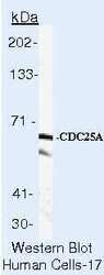
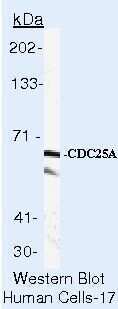
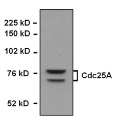
- Western blot of CDC25A using CDC25A Monoclonal Antibody (Product # MA5-13794) on LS174T Cells.
- Submitted by
- Invitrogen Antibodies (provider)
- Main image
- Experimental details
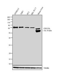
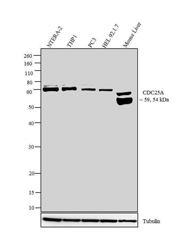
- Western blot analysis was performed on whole cell extracts (30 µg lysate) of NTERA-2 (Lane 1), THP1 (Lane 2), PC3 (Lane 3), HEL 92.1.7 (Lane 4) and Mouse Liver (Lane 5) The blots were probed with Anti-CDC25A Mouse Monoclonal Antibody (Product # MA5-13794, 1-2 µg/mL) and detected by chemiluminescence using Goat anti-Mouse IgG (H+L) Secondary Antibody, HRP conjugate (Product # 62-6520, 1:4000 dilution). Two bands ~ 59 and 54 kDa corresponding to CDC25A was observed across cell lines tested. Known quantity of protein samples were electrophoresed using Novex® NuPAGE® 12 % Bis-Tris gel (Product # NP0342BOX), XCell SureLock™ Electrophoresis System (Product # EI0002) and Novex® Sharp Pre-Stained Protein Standard (Product # LC5800). Resolved proteins were then transferred onto a nitrocellulose membrane with iBlot® 2 Dry Blotting System (Product # IB21001). The membrane was probed with the relevant primary and secondary Antibody following blocking with 5 % skimmed milk. Chemiluminescent detection was performed using Pierce™ ECL Western Blotting Substrate (Product # 32106).
- Submitted by
- Invitrogen Antibodies (provider)
- Main image
- Experimental details
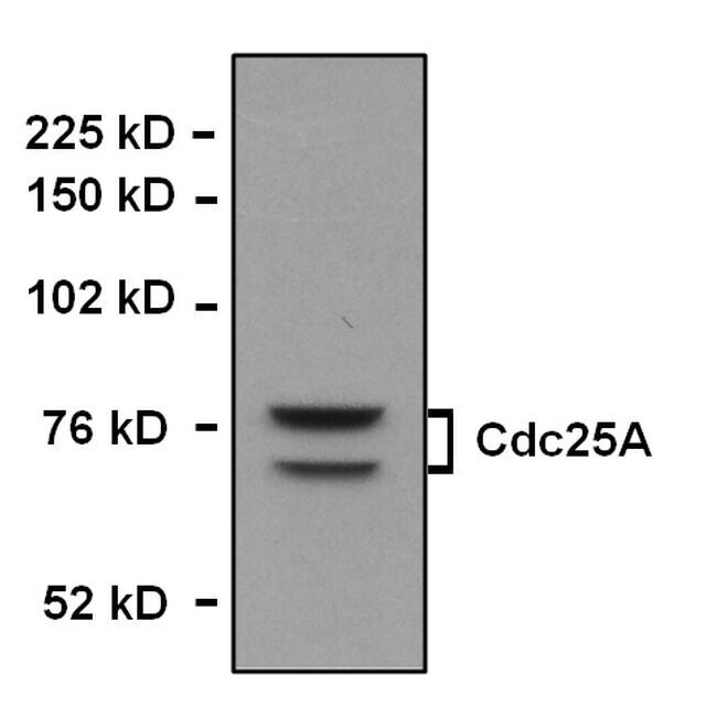
- Western blot analysis of Cdc25A was performed by loading whole cell lysate in 1X SDS sample buffer with 2-ME from 2 x 10⁵ mouse embryonic fibroblast cells. Samples were loaded onto a 4-12 % Bis-Tris polyacrylamide gel (Product # WG1402BOX). Proteins were transferred to nitrocellulose membrane with wet/tank transfer. Membrane was blocked in 5% milk/TBST. Target was detected at approximately 60 kDa using a monoclonal anti-Cdc25A antibody (Product # MA5-13794) at a dilution of 1:1000 in 5% milk/TBST at 4°C overnight, followed by a secondary antibody HRP-anti-rabbit at a dilution of 1:5000 at room temperature for 1 hour. Chemiluminescent detection was performed using Pierce™ ECL Western Blotting Substrate (Product # 32209). Data courtesy of Antibody Data Exchange Program.
Supportive validation
- Submitted by
- Invitrogen Antibodies (provider)
- Main image
- Experimental details
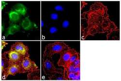
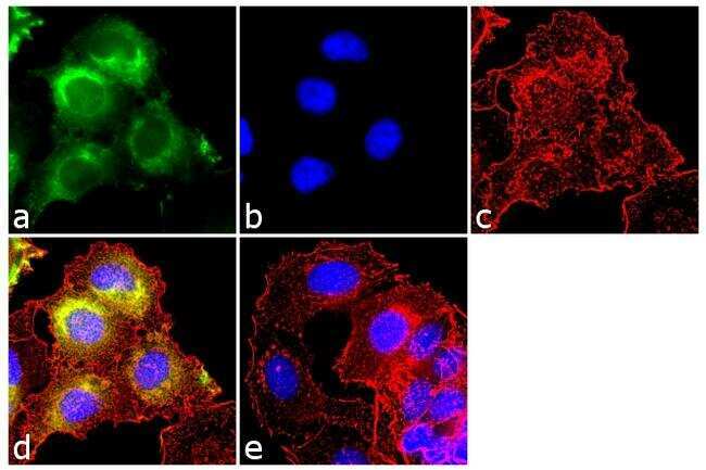
- Immunofluorescence analysis of CDC25A was done on 70% confluent log phase MCF-7 cells. The cells were fixed with 4% paraformaldehyde for 10 minutes, permeabilized with 0.1% Triton™ X-100 for 10 minutes, and blocked with 1% BSA for 1 hour at room temperature. The cells were labeled CDC25A (DCS-120) Mouse Monoclonal Antibody (Product # MA5-13794) at 2 µg/mL in 0.1% BSA and incubated for 3 hours at room temperature and then labeled with Goat anti-Mouse IgG (H+L) Superclonal™ Secondary Antibody, Alexa Fluor® 488 conjugate (Product # A28175) at a dilution of 1:2000 for 45 minutes at room temperature (Panel a: green). Nuclei (Panel b: blue) were stained with SlowFade® Gold Antifade Mountant with DAPI (Product # S36938). F-actin (Panel c: red) was stained with Alexa Fluor® 555 Rhodamine Phalloidin (Product # R415, 1:300). Panel d is a merged image showing cytoplasmic localization. Panel e is a no primary antibody control. The images were captured at 60X magnification.
- Submitted by
- Invitrogen Antibodies (provider)
- Main image
- Experimental details
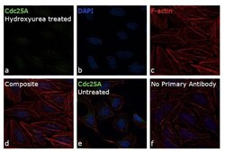
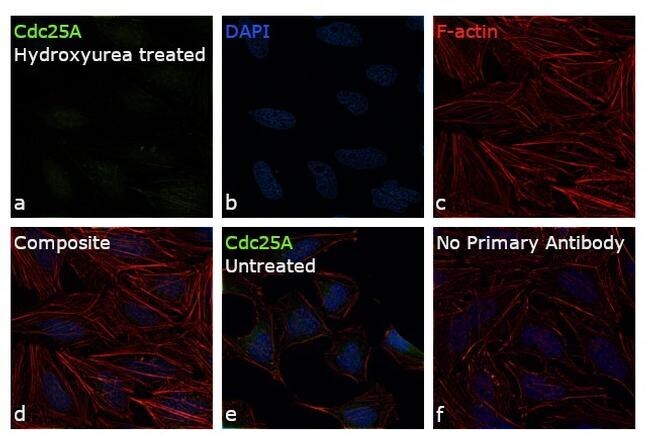
- Immunofluorescence analysis of Cdc25A was performed using 70% confluent log phase HeLa cells treated with Hydroxyurea (1mM for 10 min). The cells were fixed with 4% paraformaldehyde for 10 minutes, permeabilized with 0.1% Triton™ X-100 for 15 minutes, and blocked with 1% BSA for 1 hour at room temperature. The cells were labeled with Cdc25A Monoclonal Antibody (DCS-120) (Product # MA5-13794) at 5 µg/mL concentration in 0.1% BSA, incubated at 4 degree celsius overnight (Panel a: green). Nuclei (Panel b: blue) were stained with ProLong™ Diamond Antifade Mountant with DAPI (Product # P36962). F-actin (Panel c: red) was stained with Rhodamine Phalloidin (Product # R415, 1:300). Panel d represents the merged image showing loss of Cdc25A expression upon Hydroxyurea treatment. Panel e represents the control cells showing nuclear and cytoplasmic staining. Panel f represents control cells with no primary antibody to assess background. The images were captured at 60X magnification.
Supportive validation
- Submitted by
- Invitrogen Antibodies (provider)
- Main image
- Experimental details
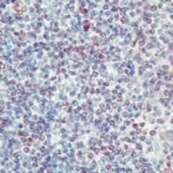
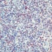
- Formalin-fixed, paraffin-embedded human tonsil stained with CDC25A antibody using peroxidase-conjugate and AEC chromogen. Note nuclear staining of epithelial cells.
Supportive validation
- Submitted by
- Invitrogen Antibodies (provider)
- Main image
- Experimental details
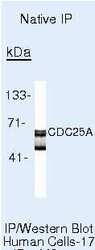
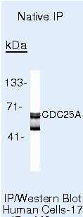
- Immunoprecipitation of CDC25A using CDC25A Monoclonal Antibody (Product # MA5-13794) on Native Human LS174T Cells.
- Submitted by
- Invitrogen Antibodies (provider)
- Main image
- Experimental details
- NULL