Explore
 Validate
 Learn
Learn Western blot
 Immunocytochemistry
Antibody data
- Antibody Data
- Antigen structure
- References [1]
- Comments [0]
- Validations
- Immunocytochemistry [1]
- Immunohistochemistry [1]
- Other assay [1]
Submit
Validation data
Reference
Comment
Report error
- Product number
- PA5-12564 - Provider product page

- Provider
- Invitrogen Antibodies
- Product name
- Phospho-Cdc25A (Thr507) Polyclonal Antibody
- Antibody type
- Polyclonal
- Antigen
- Synthetic peptide
- Description
- This antibody is predicted to react with bovine, mouse and rat based on sequence homology.
- Reactivity
- Human
- Host
- Rabbit
- Isotype
- IgG
- Vial size
- 400 μL
- Concentration
- 0.43 mg/mL
- Storage
- Store at 4°C short term. For long term storage, store at -20°C, avoiding freeze/thaw cycles.
Submitted references β-HPV 8E6 Attenuates ATM and ATR Signaling in Response to UV Damage.
Snow JA, Murthy V, Dacus D, Hu C, Wallace NA
Pathogens (Basel, Switzerland) 2019 Nov 26;8(4)
Pathogens (Basel, Switzerland) 2019 Nov 26;8(4)
No comments: Submit comment
Supportive validation
- Submitted by
- Invitrogen Antibodies (provider)
- Main image
- Experimental details
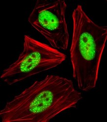
- Immunocytochemistry analysis of Phospho-Cdc25A (Thr507) in HeLa cells. Samples were incubated with Phospho-Cdc25A (Thr507) polyclonal antibody (Product # PA5-12564) using a dilution of 1:25 followed by Alexa Fluor 488-conjugated goat anti-rabbit lgG at a dilution of 1:400 (green). Cytoplasmic actin was counterstained with Alexa Fluor® 555 conjugated with Phalloidin (red).

Supportive validation
- Submitted by
- Invitrogen Antibodies (provider)
- Main image

- Experimental details
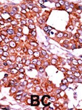
- Immunohistochemistry analysis of Phospho-Cdc25A (Thr507) in formalin-fixed and paraffin-embedded human cancer tissue. Samples were incubated with Phospho-Cdc25A (Thr507) polyclonal antibody (Product # PA5-12564) which was peroxidase-conjugated to the secondary antibody, followed by AEC staining. This data demonstrates the use of this antibody for immunohistochemistry; clinical relevance has not been evaluated. BC = breast carcinoma; HC = hepatocarcinoma.
Supportive validation
- Submitted by
- Invitrogen Antibodies (provider)
- Main image
- Experimental details
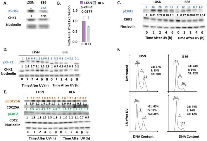
- Figure 4 beta-HPV 8E6 attenuates CHK1 phosphorylation. ( A ) Representative immunoblots of untreated hTERT HFKs with vector control (LXSN) and beta-HPV 8E6 cell lines. Nucleolin was used as a loading control. ( B ) mRNA expression level of CHEK1 in vector control (LXSN) and beta-HPV 8E6 expressing primary HFKs as measured by RT-qPCR and normalized towards the expression level of beta-actin. Data shown in figures are the means of +-SE of three independent experiments. ( C ) Representative immunoblots of hTERT HFKs with vector control (LXSN) and beta-HPV 8E6 harvested 0-6 h post 5mJ/cm 2 UVR. Nucleolin was used as a loading control. ( D ) Representative immunoblots of primary HFKs with vector control (LXSN) and beta-HPV 8E6 harvested 0-8 h post 5 mJ/cm 2 UVR. Nucleolin was used as a loading control. ( E ) Representative immunoblots of hTERT HFKs with vector control (LXSN) and beta-HPV 8E6 harvested 0-8 h post 5 mJ/cm 2 UVR. Nucleolin was used as a loading control. ( A, C - E ) The numbers above bands represent quantification by densitometry. This is shown relative to untreated cells within the same cell line and normalized to the loading control. ( F ) Cell cycle analysis of hTERT HFKs with LXSN vector control and beta-HPV 8E6 1 h post 5 mJ/cm 2 UVR.