 Explore
 Validate
 Learn
Learn Western blot
Antibody data
- Antibody Data
- Antigen structure
- References [0]
- Comments [0]
- Validations
- Western blot [2]
- Immunocytochemistry [1]
- Immunohistochemistry [2]
Submit
Validation data
Reference
Comment
Report error
- Product number
- PA5-41999 - Provider product page

- Provider
- Invitrogen Antibodies
- Product name
- FBP1 Polyclonal Antibody
- Antibody type
- Polyclonal
- Antigen
- Synthetic peptide
- Description
- Peptide sequence: YVVCFDPLDG SSNIDCLVSV GTIFGIYRKK STDEPSEKDA LQPGRNLVAA Sequence homology: Cow: 86%; Dog: 86%; Guinea Pig: 93%; Horse: 93%; Human: 100%; Mouse: 100%; Rabbit: 93%; Rat: 86%; Zebrafish: 80%
- Reactivity
- Human
- Host
- Rabbit
- Isotype
- IgG
- Vial size
- 100 µL
- Concentration
- 1 mg/mL
- Storage
- -20° C, Avoid Freeze/Thaw Cycles
No comments: Submit comment
Supportive validation
- Submitted by
- Invitrogen Antibodies (provider)
- Main image

- Experimental details
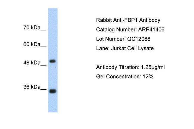
- Western blot analysis of human Jurkat cell lysate using an anti-FBP1 polyclonal antibody (Product # PA5-41999).
- Submitted by
- Invitrogen Antibodies (provider)
- Main image
- Experimental details
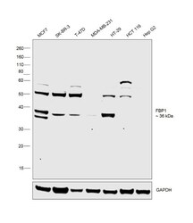
- Western blot was performed using Anti-FBP1 Polyclonal Antibody (Product # PA5-41999) and 36 kDa band corresponding to FBP1 was observed in MCF7, HT-29 and little or no expression was observed in SK-BR-3, T-47D, MDA-MB-231, HCT 116 and Hep G2. Uncharacterized bands at ~50kDa were observed across the cell lines. Whole cell extracts (30 µg lysate) of MCF7 (Lane 1), SK-BR-3 (Lane 2), T-47D (Lane 3), MDA-MB-231 (Lane 4), HT-29 (Lane 5), HCT 116 (Lane 6) and Hep G2 (Lane 7), were electrophoresed using Novex® NuPAGE® 12 % Bis-Tris gel (Product # NP0342BOX). Resolved proteins were then transferred onto a nitrocellulose membrane (Product # IB23001) by iBlot® 2 Dry Blotting System (Product # IB21001). The blot was probed with the primary antibody (1µg/mL) and detected by Goat Anti-Rabbit IgG Secondary Antibody, HRP conjugate (Product # A27036, 1:4000 dilution) using the iBright FL 1000 (Product # A32752). Chemiluminescent detection was performed using Novex® ECL Chemiluminescent Substrate Reagent Kit (Product # WP20005). .
Supportive validation
- Submitted by
- Invitrogen Antibodies (provider)
- Main image

- Experimental details
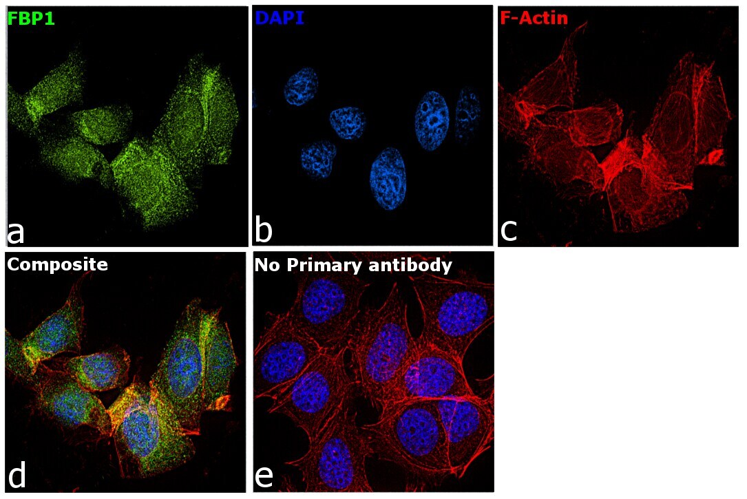
- Immunofluorescence analysis of FBP1 was performed using 70% confluent log phase MCF7 cells. The cells were fixed with 4% paraformaldehyde for 10 minutes, permeabilized with 0.1% Triton™ X-100 for 15 minutes, and blocked with 2% BSA for 1 hour at room temperature. The cells were labeled with FBP1 Polyclonal Antibody (Product # PA5-41999) at 5 microgram/mL in 0.1% BSA, incubated at 4 degree Celsius overnight and then labeled with Goat anti-Rabbit IgG (H+L) Superclonal™ Secondary Antibody, Alexa Fluor® 488 conjugate (Product # A27034) at a dilution of 1:2000 for 45 minutes at room temperature (Panel a: green). Nuclei (Panel b: blue) were stained with ProLong™ Diamond Antifade Mountant with DAPI (Product # P36962). F-actin (Panel c: red) was stained with Rhodamine Phalloidin (Product # R415, 1:300). Panel d represents the merged image showing cytosolic and nuclear localization. Panel e represents cells with no primary antibody to assess background. The images were captured at 60X magnification..
Supportive validation
- Submitted by
- Invitrogen Antibodies (provider)
- Main image
- Experimental details
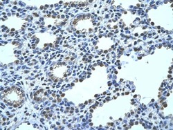
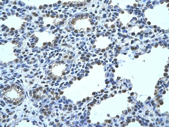
- Immunohistochemistry (paraffin-embedded) analysis of human alveolar tissue using an anti-FBP1 polyclonal antibody (Product # PA5-41999).
- Submitted by
- Invitrogen Antibodies (provider)
- Main image
- Experimental details
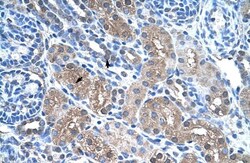
- Immunohistochemistry (paraffin-embedded) analysis of human kidney tissue using an anti-FBP1 polyclonal antibody (Product # PA5-41999).
