Explore
 Validate
 Learn
Learn Western blot
Antibody data
- Antibody Data
- Antigen structure
- References [1]
- Comments [0]
- Validations
- Western blot [3]
- Immunohistochemistry [5]
Submit
Validation data
Reference
Comment
Report error
- Product number
- NBP1-84721 - Provider product page

- Provider
- Novus Biologicals
- Proper citation
- Novus Cat#NBP1-84721, RRID:AB_11035557
- Product name
- Rabbit Polyclonal FBPase 1 Antibody
- Antibody type
- Polyclonal
- Description
- Immunogen affinity purified. Specificity of human FBPase 1 antibody verified on a Protein Array containing target protein plus 383 other non-specific proteins.
- Reactivity
- Human
- Host
- Rabbit
- Isotype
- IgG
- Vial size
- 0.1 ml
- Storage
- Store at 4C short term. Aliquot and store at -20C long term. Avoid freeze-thaw cycles.
Submitted references Fructose-1,6-bisphosphatase opposes renal carcinoma progression.
Li B, Qiu B, Lee DS, Walton ZE, Ochocki JD, Mathew LK, Mancuso A, Gade TP, Keith B, Nissim I, Simon MC
Nature 2014 Sep 11;513(7517):251-5
Nature 2014 Sep 11;513(7517):251-5
No comments: Submit comment
Supportive validation
- Submitted by
- Novus Biologicals (provider)
- Main image

- Experimental details
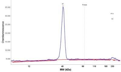
- Simple Western: FBPase 1 Antibody [NBP1-84721] - Simple Western lane view shows a specific band for FBP1 in 0.2 mg/ml of RT-4 (left) and NIH-3T3 (right) lysate. This experiment was performed under reducing conditions using the 12-230 kDa separation system.

- Submitted by
- Novus Biologicals (provider)
- Main image
- Experimental details
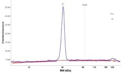
- Simple Western: FBPase 1 Antibody [NBP1-84721] - Electropherogram image(s) of corresponding Simple Western lane view. FBPase 1 antibody was used at 1:20 dilution on RT-4 and NIH-3T3 lysates(s).
- Submitted by
- Novus Biologicals (provider)
- Main image

- Experimental details
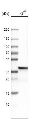
- Western Blot: FBPase 1 Antibody [NBP1-84721] - Analysis in human liver tissue.
Supportive validation
- Submitted by
- Novus Biologicals (provider)
- Main image

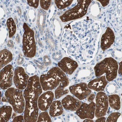
- Experimental details
- Immunohistochemistry-Paraffin: FBPase 1 Antibody [NBP1-84721] - Staining of human kidney shows moderate to strong cytoplasmic positivity in cells in tubules.
- Submitted by
- Novus Biologicals (provider)
- Main image
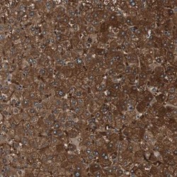
- Experimental details
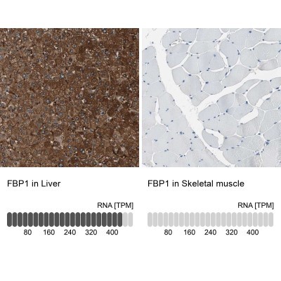
- Immunohistochemistry-Paraffin: FBPase 1 Antibody [NBP1-84721] - Staining of human liver shows moderate to strong cytoplasmic positivity in hepatocytes.
- Submitted by
- Novus Biologicals (provider)
- Main image
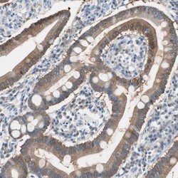
- Experimental details
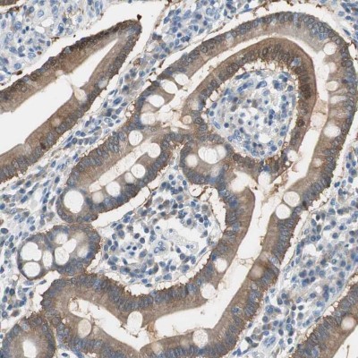
- Immunohistochemistry-Paraffin: FBPase 1 Antibody [NBP1-84721] - Staining of human duodenum shows moderate cytoplasmic positivity in glandular cells.
- Submitted by
- Novus Biologicals (provider)
- Main image
- Experimental details
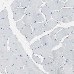
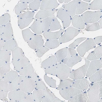
- Immunohistochemistry-Paraffin: FBPase 1 Antibody [NBP1-84721] - Staining of human skeletal muscle shows no positivity in myocytes as expected.
- Submitted by
- Novus Biologicals (provider)
- Main image
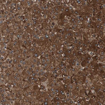
- Experimental details
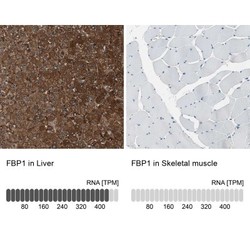
- Immunohistochemistry-Paraffin: FBPase 1 Antibody [NBP1-84721] - Staining in human liver and skeletal muscle tissues . Corresponding FBP1 RNA-seq data are presented for the same tissues.