Explore
 Validate
 Learn
Learn Western blot
 Immunohistochemistry
Antibody data
- Antibody Data
- Antigen structure
- References [10]
- Comments [0]
- Validations
- Immunohistochemistry [1]
Submit
Validation data
Reference
Comment
Report error
- Product number
- HPA005857 - Provider product page

- Provider
- Atlas Antibodies
- Proper citation
- Atlas Antibodies Cat#HPA005857, RRID:AB_1848445
- Product name
- Anti-FBP1
- Antibody type
- Polyclonal
- Description
- Polyclonal Antibody against Human FBP1, Gene description: fructose-1,6-bisphosphatase 1, Alternative Gene Names: FBP, Validated applications: WB, IHC, Uniprot ID: P09467, Storage: Store at +4°C for short term storage. Long time storage is recommended at -20°C.
- Reactivity
- Human
- Host
- Rabbit
- Conjugate
- Unconjugated
- Isotype
- IgG
- Vial size
- 100 µl
- Concentration
- 0.1 mg/ml
- Storage
- Store at +4°C for short term storage. Long time storage is recommended at -20°C.
- Handling
- The antibody solution should be gently mixed before use.
Submitted references Identification of genotype–biochemical phenotype correlations associated with fructose 1,6-bisphosphatase deficiency
Fructose-1,6-bisphosphatase 1 dephosphorylates IκBα and suppresses colorectal tumorigenesis
Extracellular vesicle-mediated communication between hepatocytes and natural killer cells promotes hepatocellular tumorigenesis
Subcellular regulation of glucose metabolism through multienzyme glucosome assemblies by EGF–ERK1/2 signaling pathways
Novel regulation of renal gluconeogenesis by Atp6ap2 in response to high fat diet via PGC1-α/AKT-1 pathway
The multi-subunit GID/CTLH E3 ubiquitin ligase promotes cell proliferation and targets the transcription factor Hbp1 for degradation
Antagonism of PPAR-γ signaling expands human hematopoietic stem and progenitor cells by enhancing glycolysis
Targeted knockdown of polo-like kinase 1 alters metabolic regulation in melanoma
Alternate Metabolic Programs Define Regional Variation of Relevant Biological Features in Renal Cell Carcinoma Progression
Fructose-1,6-bisphosphatase opposes renal carcinoma progression
Sakuma I, Nagano H, Hashimoto N, Fujimoto M, Nakayama A, Fuchigami T, Taki Y, Matsuda T, Akamine H, Kono S, Kono T, Yokoyama M, Nishimura M, Yokote K, Ogasawara T, Fujii Y, Ogawa S, Lee E, Miki T, Tanaka T
Communications Biology 2023;6(1)
Communications Biology 2023;6(1)
Fructose-1,6-bisphosphatase 1 dephosphorylates IκBα and suppresses colorectal tumorigenesis
Zhu W, Chu H, Zhang Y, Luo T, Yu H, Zhu H, Liu Y, Gao H, Zhao Y, Li Q, Wang X, Li G, Yang W
Cell Research 2023;33(3):245-257
Cell Research 2023;33(3):245-257
Extracellular vesicle-mediated communication between hepatocytes and natural killer cells promotes hepatocellular tumorigenesis
Liu Z, You Y, Chen Q, Li G, Pan W, Yang Q, Dong J, Wu Y, Bei J, Pan C, Li F, Li B
Molecular Therapy 2022;30(2):606-620
Molecular Therapy 2022;30(2):606-620
Subcellular regulation of glucose metabolism through multienzyme glucosome assemblies by EGF–ERK1/2 signaling pathways
Jeon M, Chauhan K, Szeto G, Kyoung M, An S
Journal of Biological Chemistry 2022;298(3):101675
Journal of Biological Chemistry 2022;298(3):101675
Novel regulation of renal gluconeogenesis by Atp6ap2 in response to high fat diet via PGC1-α/AKT-1 pathway
Akhtar S, Culver S, Siragy H
Scientific Reports 2021;11(1)
Scientific Reports 2021;11(1)
The multi-subunit GID/CTLH E3 ubiquitin ligase promotes cell proliferation and targets the transcription factor Hbp1 for degradation
Lampert F, Stafa D, Goga A, Soste M, Gilberto S, Olieric N, Picotti P, Stoffel M, Peter M
eLife 2018;7
eLife 2018;7
Antagonism of PPAR-γ signaling expands human hematopoietic stem and progenitor cells by enhancing glycolysis
Guo B, Huang X, Lee M, Lee S, Broxmeyer H
Nature Medicine 2018;24(3):360-367
Nature Medicine 2018;24(3):360-367
Targeted knockdown of polo-like kinase 1 alters metabolic regulation in melanoma
Gutteridge R, Singh C, Ndiaye M, Ahmad N
Cancer Letters 2017;394
Cancer Letters 2017;394
Alternate Metabolic Programs Define Regional Variation of Relevant Biological Features in Renal Cell Carcinoma Progression
Brooks S, Khandani A, Fielding J, Lin W, Sills T, Lee Y, Arreola A, Milowsky M, Wallen E, Woods M, Smith A, Nielsen M, Parker J, Lalush D, Rathmell W
Clinical Cancer Research 2016;22(12):2950-2959
Clinical Cancer Research 2016;22(12):2950-2959
Fructose-1,6-bisphosphatase opposes renal carcinoma progression
Li B, Qiu B, Lee D, Walton Z, Ochocki J, Mathew L, Mancuso A, Gade T, Keith B, Nissim I, Simon M
Nature 2014;513(7517):251-255
Nature 2014;513(7517):251-255
No comments: Submit comment
Supportive validation
- Submitted by
- Atlas Antibodies (provider)
- Enhanced method
- Orthogonal validation
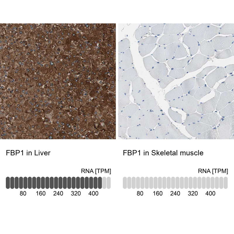
- Main image
- Experimental details
- Immunohistochemistry analysis in human liver and skeletal muscle tissues using HPA005857 antibody. Corresponding FBP1 RNA-seq data are presented for the same tissues.
- Sample type
- Human
- Protocol
- Protocol