Explore
 Validate
 Learn
Learn Western blot
 Immunohistochemistry
Antibody data
- Antibody Data
- Antigen structure
- References [0]
- Comments [0]
- Validations
- Western blot [1]
- Flow cytometry [1]
Submit
Validation data
Reference
Comment
Report error
- Product number
- PA1-602 - Provider product page

- Provider
- Invitrogen Antibodies
- Product name
- TEM8 Polyclonal Antibody
- Antibody type
- Polyclonal
- Antigen
- Synthetic peptide
- Description
- PA1-602 detects Tem-8 from mouse samples. PA1-602 has been successfully used in Western blot and immunohistochemical procedures. By Western blot, this antibody detects ~45 kDa, 60 kDa, 85 kDa proteins, representing Tem-8 from mouse tissue extracts.This antibody has also been used in immunohistochemical procedures (paraffin samples) using mouse tissues. PA1-602 immunizing peptide corresponds to amino acid residues 92-107, within the extracellular domain, of human ATR/TEM8. This peptide (Cat. # PEP-253) is available for neutralization and control experiments.
- Reactivity
- Human, Mouse
- Host
- Rabbit
- Isotype
- IgG
- Vial size
- 100 μg
- Concentration
- 1 mg/mL
- Storage
- -20°C, Avoid Freeze/Thaw Cycles
No comments: Submit comment
Supportive validation
- Submitted by
- Invitrogen Antibodies (provider)
- Main image
- Experimental details
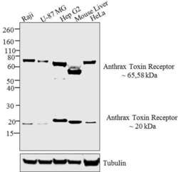
- Western blot analysis was performed using membrane enriched extracts (30 µg) of Raji (Lane 1), U-87 MG (Lane 2), Hep G2 (Lane 3), Mouse Liver (Lane 4) and HeLa (Lane 5).The blots were probed with Anti-Anthrax Toxin Receptor Rabbit Polyclonal Antibody (Product # PA1-602, 1 in 1000 dilution) and detected by chemiluminescence using Goat anti-Rabbit IgG (Heavy Chain) Superclonal™ Secondary Antibody, HRP conjugate (Product # A27036, 0.4µg/mL 1:2500 dilution). A~ 65kDa band of Anthrax Toxin Receptor was observed across the cell lines tested. A ~58kDa band was observed in the Mouse liver tissue sample. A band of 20 kDa was also observed across the panel of cell lines and tissues tested. This could be due to the separation of the PA subunit, of the Anthrax Toxin Receptor, into two subunits of 65kDa and 20kDa, upon trypsinolysis. A Known quantity of protein samples were electrophoresed using Novex® NuPAGE®12 % Bis-Tris gel (Product # NP0342BOX), XCell SureLock™ Electrophoresis System (Product # EI0002) and Novex® Sharp Pre-Stained Protein Standard (Product # LC5800). Resolved proteins were then transferred onto a nitrocellulose membrane by iBlot® 2 Dry Blotting System (Product # IB21001). The membrane was probed with the relevant primary and secondary Antibody following blocking with 5 % skimmed milk. Chemiluminescent detection was performed using Pierce™ ECL Western Blotting Substrate (Product # 32106).

Supportive validation
- Submitted by
- Invitrogen Antibodies (provider)
- Main image
- Experimental details
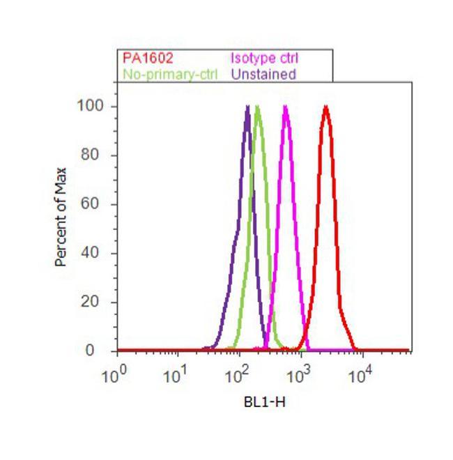
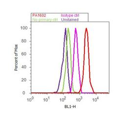
- Flow cytometry analysis of Anthrax Toxin Receptor was performed using SH-SY5Y cells. Cells were fixed with 70% ethanol for 10 minutes, permeabilized with 0.25% Triton™ X-100 for 20 minutes, and blocked with 5% BSA for 30 minutes at room temperature. Cells were labeled with Anthrax Toxin Receptor Rabbit Polyclonal Antibody (Product # PA1-602, red histogram) or with rabbit isotype control (pink histogram) at 3-5 µg/million cells in 2.5% BSA. After incubation at room temperature for 2 hours, the cells were labeled with Alexa Fluor® 488 Goat Anti-Rabbit Secondary Antibody (Product # A11008) at a dilution of 1:400 for 30 minutes at room temperature. The representative 10, 000 cells were acquired and analyzed for each sample using an Attune® Acoustic Focusing Cytometer. The purple histogram represents unstained control cells and the green histogram represents no-primary-antibody control.