 Explore
 Validate
 Learn
Learn Western blot
Antibody data
- Antibody Data
- Antigen structure
- References [1]
- Comments [0]
- Validations
- Western blot [3]
- Immunohistochemistry [2]
Submit
Validation data
Reference
Comment
Report error
- Product number
- GTX129241 - Provider product page

- Provider
- GeneTex
- Proper citation
- GeneTex Cat#GTX129241, RRID:AB_2783558
- Product name
- LHX2 antibody
- Antibody type
- Polyclonal
- Reactivity
- Human, Mouse, Rat
- Host
- Rabbit
Submitted references A cell identity switch allows residual BCC to survive Hedgehog pathway inhibition.
Biehs B, Dijkgraaf GJP, Piskol R, Alicke B, Boumahdi S, Peale F, Gould SE, de Sauvage FJ
Nature 2018 Oct;562(7727):429-433
Nature 2018 Oct;562(7727):429-433
No comments: Submit comment
Supportive validation
- Submitted by
- GeneTex (provider)
- Main image

- Experimental details
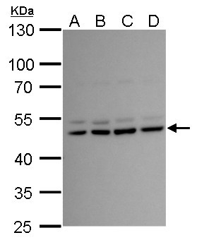
- LHX2 antibody detects LHX2 protein by western blot analysis.A 293T whole cell lysate/extract B A431 whole cell lysate/extractC HeLa D HepG2 whole cell lysate/extract10% SDS-PAGELHX2 antibody (GTX129241) dilution: 1:1000 The HRP-conjugated anti-rabbit IgG antibody (GTX213110-01) was used to detect the primary antibody.
- Submitted by
- GeneTex (provider)
- Main image
- Experimental details
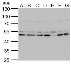
- LHX2 antibody detects LHX2 protein by western blot analysis.A. Neuro2A whole cell lysate/extractB. GL261 whole cell lysate/extractC. C8D30 whole cell lysate/extractD. NIH-3T3 whole cell lysate/extractE. BCL-1 whole cell lysate/extractF. BCL-1 whole cell lysate/extractG. Raw264.7 whole cell lysate/extractH. C2C12 whole cell lysate/extract10% SDS-PAGELHX2 antibody (GTX129241) dilution: 1:1000 The HRP-conjugated anti-rabbit IgG antibody (GTX213110-01) was used to detect the primary antibody.

- Submitted by
- GeneTex (provider)
- Main image
- Experimental details
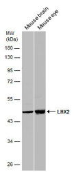
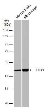
- Various tissue extracts (50 ?g) were separated by 10% SDS-PAGE, and the membranes were blotted with LHX2 antibody (GTX129241) diluted at 1:10000 (left panel) and 1:5000 (right panel). The HRP-conjugated anti-rabbit IgG antibody (GTX213110-01) was used to detect the primary antibody.
Supportive validation
- Submitted by
- GeneTex (provider)
- Main image
- Experimental details
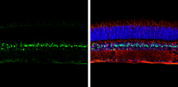
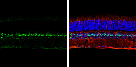
- LHX2 antibody detects LHX2 protein expression by immunohistochemical analysis.Sample: Frozen sectioned adult mouse retina. Green: LHX2 protein stained by LHX2 antibody (GTX129241) diluted at 1:250.Red: beta Tubulin 3/ TUJ1, stained by beta Tubulin 3/ TUJ1 antibody [GT11710] (GTX631836) diluted at 1:250.Blue: Fluoroshield with DAPI (GTX30920).
- Submitted by
- GeneTex (provider)
- Main image
- Experimental details
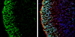
- LHX2 antibody detects LHX2 protein by immunohistochemical analysis.Sample: Frozen-sectioned rat E13.5 brain.Green: LHX2 stained by LHX2 antibody (GTX129241) diluted at 1:250.Red: beta Tubulin 3/ Tuj1, stained by beta Tubulin 3/ Tuj1 antibody [GT1338] (GTX631831) diluted at 1:500.Blue: Fluoroshield with DAPI (GTX30920).
