Explore
 Validate
 Learn
Learn Western blot
 Immunohistochemistry
Antibody data
- Antibody Data
- Antigen structure
- References [1]
- Comments [0]
- Validations
- Immunohistochemistry [2]
- Other assay [1]
Submit
Validation data
Reference
Comment
Report error
- Product number
- PA5-78287 - Provider product page

- Provider
- Invitrogen Antibodies
- Product name
- LHX2 Polyclonal Antibody
- Antibody type
- Polyclonal
- Antigen
- Recombinant full-length protein
- Description
- Positive Control: 293T, A431, HeLa, HepG2, Neuro 2A, GL261, C8D30, NIH-3T3, BCL-1, Raw264.7, C2C12, mouse brain, mouse eye Predicted Reactivity: Pig (98%), Chicken (81%), Rhesus Monkey (98%), Bovine (98%) Store product as a concentrated solution. Centrifuge briefly prior to opening the vial.
- Reactivity
- Human, Mouse, Rat
- Host
- Rabbit
- Isotype
- IgG
- Vial size
- 100 μL
- Concentration
- 0.97 mg/mL
- Storage
- Store at 4°C short term. For long term storage, store at -20°C, avoiding freeze/thaw cycles.
Submitted references Chromosome 10q26-driven age-related macular degeneration is associated with reduced levels of HTRA1 in human retinal pigment epithelium.
Williams BL, Seager NA, Gardiner JD, Pappas CM, Cronin MC, Amat di San Filippo C, Anstadt RA, Liu J, Toso MA, Nichols L, Parnell TJ, Eve JR, Heinz S, Hayes MGB, Bartel PL, Zouache MA, Richards BT, Hageman GS
Proceedings of the National Academy of Sciences of the United States of America 2021 Jul 27;118(30)
Proceedings of the National Academy of Sciences of the United States of America 2021 Jul 27;118(30)
No comments: Submit comment
Supportive validation
- Submitted by
- Invitrogen Antibodies (provider)
- Main image

- Experimental details
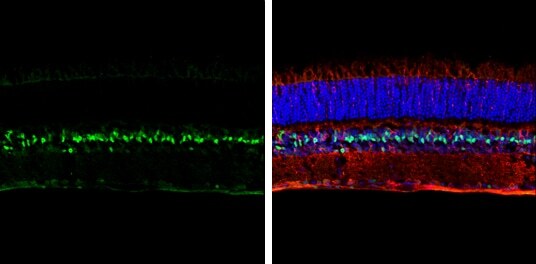
- LHX2 Polyclonal Antibody detects LHX2 protein expression by immunohistochemical analysis. Sample: Frozen sectioned adult mouse retina. Green: LHX2 protein stained by LHX2 Polyclonal Antibody (Product # PA5-78287) diluted at 1:250. Red: beta Tubulin 3/ TUJ1, stained by beta Tubulin 3/ TUJ1 antibody [GT11710] diluted at 1:250. Blue: Fluoroshield with DAPI .
- Submitted by
- Invitrogen Antibodies (provider)
- Main image
- Experimental details
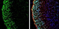
- Immunohistochemistry (Frozen) analysis of LHX2 was performed in frozen-sectioned rat E13.5 brain tissue using LHX2 Polyclonal Antibody (Product # PA5-78287) at a dilution of 1:250 (Green). Red: beta Tubulin 3/ Tuj1, stained by beta Tubulin 3/ Tuj1 antibody diluted at 1:500. Blue: Fluoroshield with DAPI.

Supportive validation
- Submitted by
- Invitrogen Antibodies (provider)
- Main image
- Experimental details
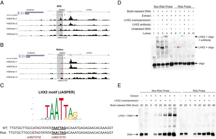
- Fig. 4. Epigenetic analysis of Chr10 AMD-associated locus. ( A and B ) ChIP-Seq analysis of H3K27Ac and H3K4me1 in adult human RPE ( A ) or retina ( B ) tissue from two independent homozygous nonrisk donors (-1 and -2). Normalized fragment coverage is displayed. The gray region represents the 4-kb HTRA1 regulatory region, and the red line marks the location of rs36212733. ( C ) Consensus Lhx2 DNA binding motif and predicted Lhx2 binding site (underlined) within the 4-kb HTRA1 regulatory region on Chr10 overlapping the rs36212733 SNP. ( D ) EMSA analysis for SNP-genotype-specific binding of nuclear extracts from HEK293 cells with (+) or without (-) transient Lhx2 overexpression to oligonucleotide (oligo) probes with the risk or nonrisk SNPs at rs36212732 and rs36212733. Anti-Lhx2 antibody or unlabeled probe was included in some reactions, as indicated. Red asterisk indicates band found in Lhx2-containing nuclear extracts but not extracts without Lhx2 overexpression. Green asterisk indicates the band supershifted with addition of Lhx2 antibody to the reaction. ( E ) Same as in D , except that increasing amounts of biotin-labeled probe were included in the reaction mixture.