Explore
 Validate
 Learn
LearnA02636-1
antibody from Boster Biological Technology
Targeting: PCBP1
hnRNP-E1, hnRNP-X, HNRPE1, HNRPX
 Western blot
 ELISA
ELISAAntibody data
- Antibody Data
- Antigen structure
- References [1]
- Comments [0]
- Validations
- Western blot [1]
Submit
Validation data
Reference
Comment
Report error
- Product number
- A02636-1 - Provider product page

- Provider
- Boster Biological Technology
- Product name
- Anti-PCBP1 Antibody Picoband™
- Antibody type
- Polyclonal
- Description
- Rabbit IgG polyclonal antibody for PCBP1 detection. Tested with WB, IHC-P, FCM, Direct ELISA in Human;Mouse;Rat.
- Reactivity
- Human, Mouse, Rat
- Host
- Rabbit
- Vial size
- 100μg/vial
- Concentration
- Add 0.2ml of distilled water will yield a concentration of 500μg/ml.
- Storage
- At -20°C for one year. After reconstitution, at 4°C for one month. It can also be aliquoted and stored frozen at -20°C for a longer time. Avoid repeated freezing and thawing.
- Handling
- Add 0.2ml of distilled water will yield a concentration of 500μg/ml.
Submitted references Generation of PCBP1-deficient pigs using CRISPR/Cas9-mediated gene editing.
Qi C, Pang D, Yang K, Jiao S, Wu H, Zhao C, Hu L, Li F, Zhou J, Yang L, Lv D, Tang X, Ouyang H, Xie Z
iScience 2022 Oct 21;25(10):105268
iScience 2022 Oct 21;25(10):105268
No comments: Submit comment
Supportive validation
- Submitted by
- Boster Biological Technology (provider)
- Main image
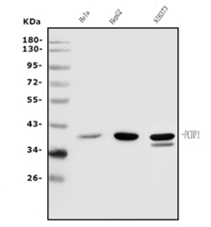
- Experimental details
- Western blot analysis of PCBP1 using anti-PCBP1 antibody (A02636-1). Electrophoresis was performed on a 5-20% SDS-PAGE gel at 70V (Stacking gel) / 90V (Resolving gel) for 2-3 hours. The sample well of each lane was loaded with 50ug of sample under reducing conditions. Lane 1: human HELA whole cell lysates, Lane 2: human HEPG2 whole cell lysates, Lane 3: mouse NIH/3T3 whole cell lysates. After Electrophoresis, proteins were transferred to a Nitrocellulose membrane at 150mA for 50-90 minutes. Blocked the membrane with 5% Non-fat Milk/ TBS for 1.5 hour at RT. The membrane was incubated with rabbit anti-PCBP1 antigen affinity purified polyclonal antibody (Catalog # A02636-1) at 0.5 μg/mL overnight at 4°C, then washed with TBS-0.1%Tween 3 times with 5 minutes each and probed with a goat anti-rabbit IgG-HRP secondary antibody at a dilution of 1:5000 for 1.5 hour at RT. The signal is developed using an Enhanced Chemiluminescent detection (ECL) kit (Catalog # EK1002) with Tanon 5200 system. A specific band was detected for PCBP1 at approximately 40KD. The expected band size for PCBP1 is at 40KD.
- Additional image