Explore
 Validate
 Learn
Learn Western blot
Antibody data
- Antibody Data
- Antigen structure
- References [3]
- Comments [0]
- Validations
- Western blot [2]
- Immunohistochemistry [1]
Submit
Validation data
Reference
Comment
Report error
- Product number
- AF3355 - Provider product page

- Provider
- R&D Systems
- Product name
- Mouse/Rat CD200 Antibody
- Antibody type
- Polyclonal
- Description
- Antigen Affinity-purified. Detects mouse CD200 in direct ELISAs and mouse CD200 and rat CD200 Western blots. In direct ELISAs, approximately 50% cross-reactivity with recombinant human CD200 is observed.
- Reactivity
- Mouse, Rat
- Host
- Goat
- Conjugate
- Unconjugated
- Antigen sequence
O54901- Isotype
- IgG
- Vial size
- 100 ug
- Concentration
- LYOPH
- Storage
- Use a manual defrost freezer and avoid repeated freeze-thaw cycles. 12 months from date of receipt, -20 to -70 °C as supplied. 1 month, 2 to 8 °C under sterile conditions after reconstitution. 6 months, -20 to -70 °C under sterile conditions after reconstitution.
Submitted references Dok2 mediates the CD200Fc attenuation of Aβ-induced changes in glia.
Bald scalp in men with androgenetic alopecia retains hair follicle stem cells but lacks CD200-rich and CD34-positive hair follicle progenitor cells.
Potent immunosuppression by a bivalent molecule binding to CD200R and TGF-betaR.
Lyons A, Downer EJ, Costello DA, Murphy N, Lynch MA
Journal of neuroinflammation 2012 May 29;9:107
Journal of neuroinflammation 2012 May 29;9:107
Bald scalp in men with androgenetic alopecia retains hair follicle stem cells but lacks CD200-rich and CD34-positive hair follicle progenitor cells.
Garza LA, Yang CC, Zhao T, Blatt HB, Lee M, He H, Stanton DC, Carrasco L, Spiegel JH, Tobias JW, Cotsarelis G
The Journal of clinical investigation 2011 Feb;121(2):613-22
The Journal of clinical investigation 2011 Feb;121(2):613-22
Potent immunosuppression by a bivalent molecule binding to CD200R and TGF-betaR.
Gorczynski RM, Chen Z, Shivagnahnam S, Taseva A, Wong K, Yu K, Khatri I
Transplantation 2010 Jul 27;90(2):150-9
Transplantation 2010 Jul 27;90(2):150-9
No comments: Submit comment
Supportive validation
- Submitted by
- R&D Systems (provider)
- Main image

- Experimental details
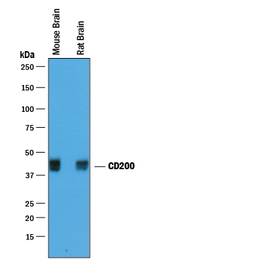
- Detection of Mouse and Rat CD200 by Western Blot. Western blot shows lysates of mouse brain tissue and rat brain tissue. PVDF membrane was probed with 0.25 µg/mL of Goat Anti-Mouse/Rat CD200 Antigen Affinity-purified Polyclonal Antibody (Catalog # AF3355) followed by HRP-conjugated Anti-Goat IgG Secondary Antibody (Catalog # HAF019). A specific band was detected for CD200 at approximately 38-45 kDa (as indicated). This experiment was conducted under reducing conditions and using Immunoblot Buffer Group 1.
- Submitted by
- R&D Systems (provider)
- Main image
- Experimental details
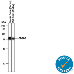
- Detection of Mouse and Rat CD200 by Simple WesternTM. Simple Western lane view shows lysates of mouse brain (cortex) tissue and rat brain (cortex) tissue, loaded at 0.2 mg/mL. A specific band was detected for CD200 at approximately 73-76 kDa (as indicated) using 2.5 µg/mL of Goat Anti-Mouse/Rat CD200 Antigen Affinity-purified Polyclonal Antibody (Catalog # AF3355) followed by 1:50 dilution of HRP-conjugated Anti-Goat IgG Secondary Antibody (Catalog # HAF109). This experiment was conducted under reducing conditions and using the 12-230 kDa separation system.

Supportive validation
- Submitted by
- R&D Systems (provider)
- Main image
- Experimental details
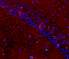
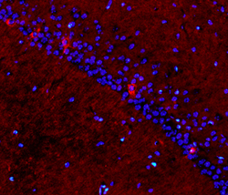
- CD200 in Mouse Brain. CD200 was detected in perfusion fixed frozen sections of normal mouse brain using Goat Anti-Mouse/Rat CD200 Antigen Affinity-purified Polyclonal Antibody (Catalog # AF3355) at 15 µg/mL overnight at 4 °C. Tissue was stained using the NorthernLights™ 557-conjugated Anti-Goat IgG Secondary Antibody (red; Catalog # NL001) and counterstained with DAPI (blue). Specific staining was localized to plasma membranes of hippocampal neurons. View our protocol for Fluorescent IHC Staining of Frozen Tissue Sections.