Explore
 Validate
 Learn
Learn Immunohistochemistry
 Flow cytometry
Antibody data
- Antibody Data
- Antigen structure
- References [6]
- Comments [0]
- Validations
- Flow cytometry [2]
- Other assay [5]
Submit
Validation data
Reference
Comment
Report error
- Product number
- 14-9200-80 - Provider product page

- Provider
- Invitrogen Antibodies
- Product name
- CD200 Monoclonal Antibody (OX104), eBioscience™
- Antibody type
- Monoclonal
- Antigen
- Other
- Description
- Description: The monoclonal antibody OX104 recognizes human CD200 also known as OX2. CD200 is a member of the Ig superfamily with 2 Ig domains, a transmembrane and cytoplasmic domain. CD200 is expressed on resting and activated B cells, a subset of resting and activated T cells, keratinocytes, peripheral and central nerve cells, follicular dendritic cells and ovarian cells. The interaction with CD200R results in macrophage activation (IL-6 production), inhibition of mast cell degranulation along with reduced TNFalpha and IL-13 secretion and overall attenuation of the activation status of lymphocytes. A role has also been suggested in maternal tolerance as expression of CD200 is also present on the trophoblast. Applications Reported: This OX104 antibody has been reported for use in flow cytometric analysis, and immunohistology staining of frozen tissue sections. Applications Tested: This OX104 antibody has been tested by flow cytometric analysis of normal human peripheral blood cells. This can be used at less than or equal to 0.25 µg per test. A test is defined as the amount (µg) of antibody that will stain a cell sample in a final volume of 100 µL. Cell number should be determined empirically but can range from 10^5 to 10^8 cells/test. It is recommended that the antibody be carefully titrated for optimal performance in the assay of interest. Purity: Greater than 90%, as determined by SDS-PAGE. Aggregation: Less than 10%, as determined by HPLC. Filtration: 0.2 µm post-manufacturing filtered.
- Reactivity
- Human
- Host
- Mouse
- Isotype
- IgG
- Antibody clone number
- OX104
- Vial size
- 25 μg
- Concentration
- 0.5 mg/mL
- Storage
- 4°C
Submitted references A Critical Role for CD200R Signaling in Limiting the Growth and Metastasis of CD200+ Melanoma.
Residual malignant and normal plasma cells shortly after high dose melphalan and stem cell transplantation. Highlight of a putative therapeutic window in Multiple Myeloma?
Analysis of leukocyte membrane protein interactions using protein microarrays.
Human herpesvirus 8 K14 protein mimics CD200 in down-regulating macrophage activation through CD200 receptor.
Characterization of the CD200 receptor family in mice and humans and their interactions with CD200.
The unusual distribution of the neuronal/lymphoid cell surface CD200 (OX2) glycoprotein is conserved in humans.
Liu JQ, Talebian F, Wu L, Liu Z, Li MS, Wu L, Zhu J, Markowitz J, Carson WE 3rd, Basu S, Bai XF
Journal of immunology (Baltimore, Md. : 1950) 2016 Aug 15;197(4):1489-97
Journal of immunology (Baltimore, Md. : 1950) 2016 Aug 15;197(4):1489-97
Residual malignant and normal plasma cells shortly after high dose melphalan and stem cell transplantation. Highlight of a putative therapeutic window in Multiple Myeloma?
Caraux A, Vincent L, Bouhya S, Quittet P, Moreaux J, Requirand G, Veyrune JL, Olivier G, Cartron G, Rossi JF, Klein B
Oncotarget 2012 Nov;3(11):1335-47
Oncotarget 2012 Nov;3(11):1335-47
Analysis of leukocyte membrane protein interactions using protein microarrays.
Letarte M, Voulgaraki D, Hatherley D, Foster-Cuevas M, Saunders NJ, Barclay AN
BMC biochemistry 2005 Mar 1;6:2
BMC biochemistry 2005 Mar 1;6:2
Human herpesvirus 8 K14 protein mimics CD200 in down-regulating macrophage activation through CD200 receptor.
Foster-Cuevas M, Wright GJ, Puklavec MJ, Brown MH, Barclay AN
Journal of virology 2004 Jul;78(14):7667-76
Journal of virology 2004 Jul;78(14):7667-76
Characterization of the CD200 receptor family in mice and humans and their interactions with CD200.
Wright GJ, Cherwinski H, Foster-Cuevas M, Brooke G, Puklavec MJ, Bigler M, Song Y, Jenmalm M, Gorman D, McClanahan T, Liu MR, Brown MH, Sedgwick JD, Phillips JH, Barclay AN
Journal of immunology (Baltimore, Md. : 1950) 2003 Sep 15;171(6):3034-46
Journal of immunology (Baltimore, Md. : 1950) 2003 Sep 15;171(6):3034-46
The unusual distribution of the neuronal/lymphoid cell surface CD200 (OX2) glycoprotein is conserved in humans.
Wright GJ, Jones M, Puklavec MJ, Brown MH, Barclay AN
Immunology 2001 Feb;102(2):173-9
Immunology 2001 Feb;102(2):173-9
No comments: Submit comment
Supportive validation
- Submitted by
- Invitrogen Antibodies (provider)
- Main image

- Experimental details
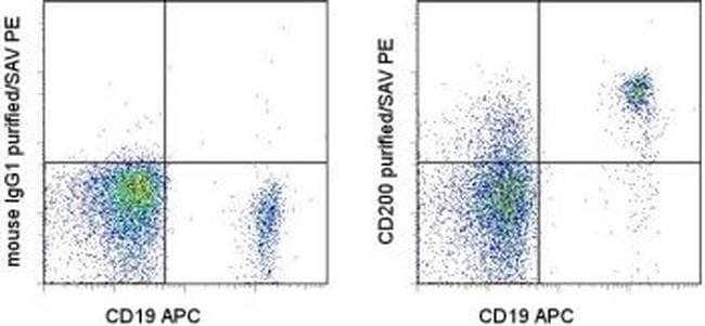
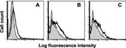
- Staining of normal human peripheral blood cells with Anti-Human CD19 APC (Product # 17-0199-42) and 0.125 µg of Mouse IgG1 kappa Isotype Control Purified (Product # 14-4714-82) (left) or 0.125 µg of Anti-Human CD200 Purified (right) followed by Anti-Mouse IgG Biotin (Product # 13-4013-85) and Streptavidin PE (Product # 12-4317-87). Cells in the lymphocyte gate were used for analysis.
- Submitted by
- Invitrogen Antibodies (provider)
- Main image

- Experimental details
- Staining of normal human peripheral blood cells with Anti-Human CD19 APC (Product # 17-0199-42) and 0.125 µg of Mouse IgG1 kappa Isotype Control Purified (Product # 14-4714-82) (left) or 0.125 µg of Anti-Human CD200 Purified (right) followed by Anti-Mouse IgG Biotin (Product # 13-4013-85) and Streptavidin PE (Product # 12-4317-87). Cells in the lymphocyte gate were used for analysis.

Supportive validation
- Submitted by
- Invitrogen Antibodies (provider)
- Main image

- Experimental details
- NULL
- Submitted by
- Invitrogen Antibodies (provider)
- Main image
- Experimental details
- NULL

- Submitted by
- Invitrogen Antibodies (provider)
- Main image
- Experimental details
- NULL

- Submitted by
- Invitrogen Antibodies (provider)
- Main image
- Experimental details
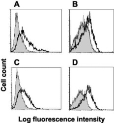
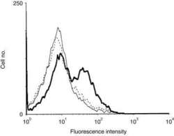
- 1 Recombinant human OX2CD4d3+4 binds rat OX2 receptor (OX2R). Flow cytometry analysis shows that human OX2-coated beads bound rat resident peritoneal cells (RPCs) coated in OX45 (anti-rat CD48) monoclonal antibody (mAb) (thick line), but that binding was blocked back to a negative control (CD4d3+4-coated beads, thin line) by OX102 (anti-rat OX2R) mAb (broken line).
- Submitted by
- Invitrogen Antibodies (provider)
- Main image

- Experimental details
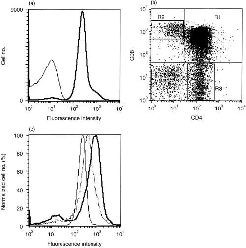
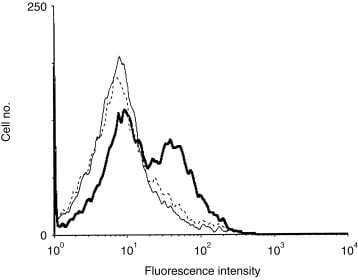
- 3 OX2 is expressed on thymocytes and at increased levels on single positive, lineage-committed thymocytes. (a) Flow cytometry shows that almost all thymocytes were stained with the OX104 monoclonal antibody (mAb) (thick line) above an isotype-matched negative control, OX1 (thin line). (b) Thymocytes were gated, according to CD4 and CD8 expression, into: (R1) CD4 + CD8 + double-positives (DPs); (R2) CD8 + single-positives (SPs); and (R3) CD4 + SPs. The level of OX2 expression was plotted as a function of the normalized cell number from each region in (b) with R3 CD4 + SPs (bold line), R2 CD8 + SPs (dotted line) and R1 CD4 + CD8 + DPs (thin line) (c).