 Explore
 Validate
 Learn
Learn Western blot
 Immunocytochemistry
 Immunoprecipitation
Antibody data
- Antibody Data
- Antigen structure
- References [0]
- Comments [0]
- Validations
- Immunocytochemistry [4]
- Immunohistochemistry [4]
- Other assay [1]
Submit
Validation data
Reference
Comment
Report error
- Product number
- MA5-32615 - Provider product page

- Provider
- Invitrogen Antibodies
- Product name
- TIA-1 Recombinant Rabbit Monoclonal Antibody (JM42-11)
- Antibody type
- Monoclonal
- Antigen
- Synthetic peptide
- Description
- Recombinant rabbit monoclonal antibodies are produced using in vitro expression systems. The expression systems are developed by cloning in the specific antibody DNA sequences from immunoreactive rabbits. Then, individual clones are screened to select the best candidates for production. The advantages of using recombinant rabbit monoclonal antibodies include: better specificity and sensitivity, lot-to-lot consistency, animal origin-free formulations, and broader immunoreactivity to diverse targets due to larger rabbit immune repertoire.
- Reactivity
- Human, Mouse
- Host
- Rabbit
- Isotype
- IgG
- Antibody clone number
- JM42-11
- Vial size
- 100 μL
- Concentration
- 1 mg/mL
- Storage
- Store at 4°C short term. For long term storage, store at -20°C, avoiding freeze/thaw cycles.
No comments: Submit comment
Supportive validation
- Submitted by
- Invitrogen Antibodies (provider)
- Main image
- Experimental details
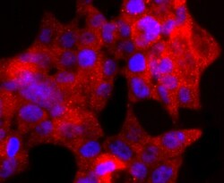
- Immunocytochemical analysis of TIA-1 in 293T cells using a TIA-1 Monoclonal antibody (Product # MA5-32615) as seen in red. The nuclear counter stain is DAPI (blue). Cells were fixed in paraformaldehyde, permeabilised with 0.25% Triton X100/PBS.

- Submitted by
- Invitrogen Antibodies (provider)
- Main image
- Experimental details
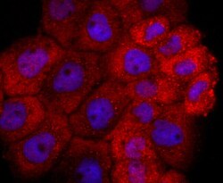
- Immunocytochemical analysis of TIA-1 in A431 cells using a TIA-1 Monoclonal antibody (Product # MA5-32615) as seen in red. The nuclear counter stain is DAPI (blue). Cells were fixed in paraformaldehyde, permeabilised with 0.25% Triton X100/PBS.
- Submitted by
- Invitrogen Antibodies (provider)
- Main image

- Experimental details
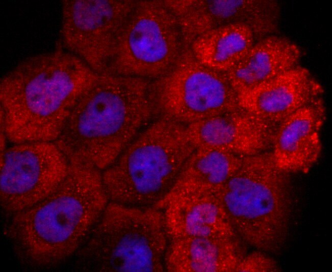
- Immunocytochemical analysis of TIA-1 in Hela cells using a TIA-1 Monoclonal antibody (Product # MA5-32615) as seen in red. The nuclear counter stain is DAPI (blue). Cells were fixed in paraformaldehyde, permeabilised with 0.25% Triton X100/PBS.
- Submitted by
- Invitrogen Antibodies (provider)
- Main image
- Experimental details
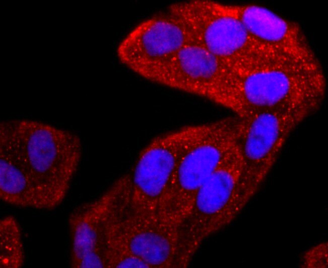
- Immunocytochemistry analysis of TIA-1 with Hela cells (red). Formalin fixed cells were permeabilized with 0.1% Triton X-100 in TBS (10 min, room temp) and blocked with 1% Blocker BSA (15 min, room temp), incubated with recombinant monoclonal TIA-1 (Product # MA5-32615) at a dilution of 1:50 for 1 hour at room temperature, washed with PBS, followed by Alexa Fluor 594 Goat anti-Rabbit IgG at 1:1,000 dilution. DAPI was used to stain the cell nuclei (blue).
Supportive validation
- Submitted by
- Invitrogen Antibodies (provider)
- Main image

- Experimental details
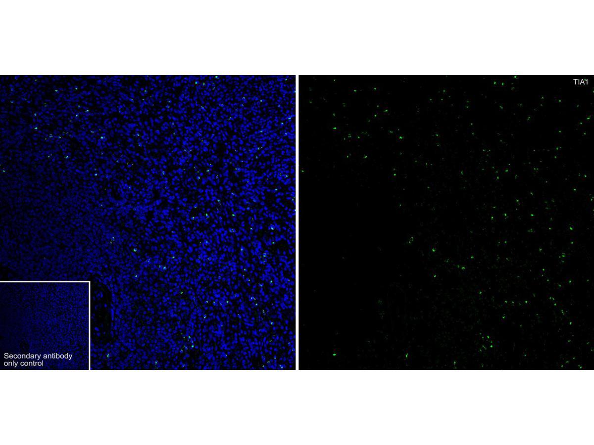
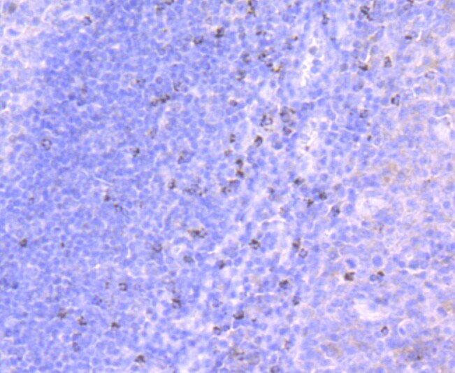
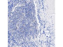
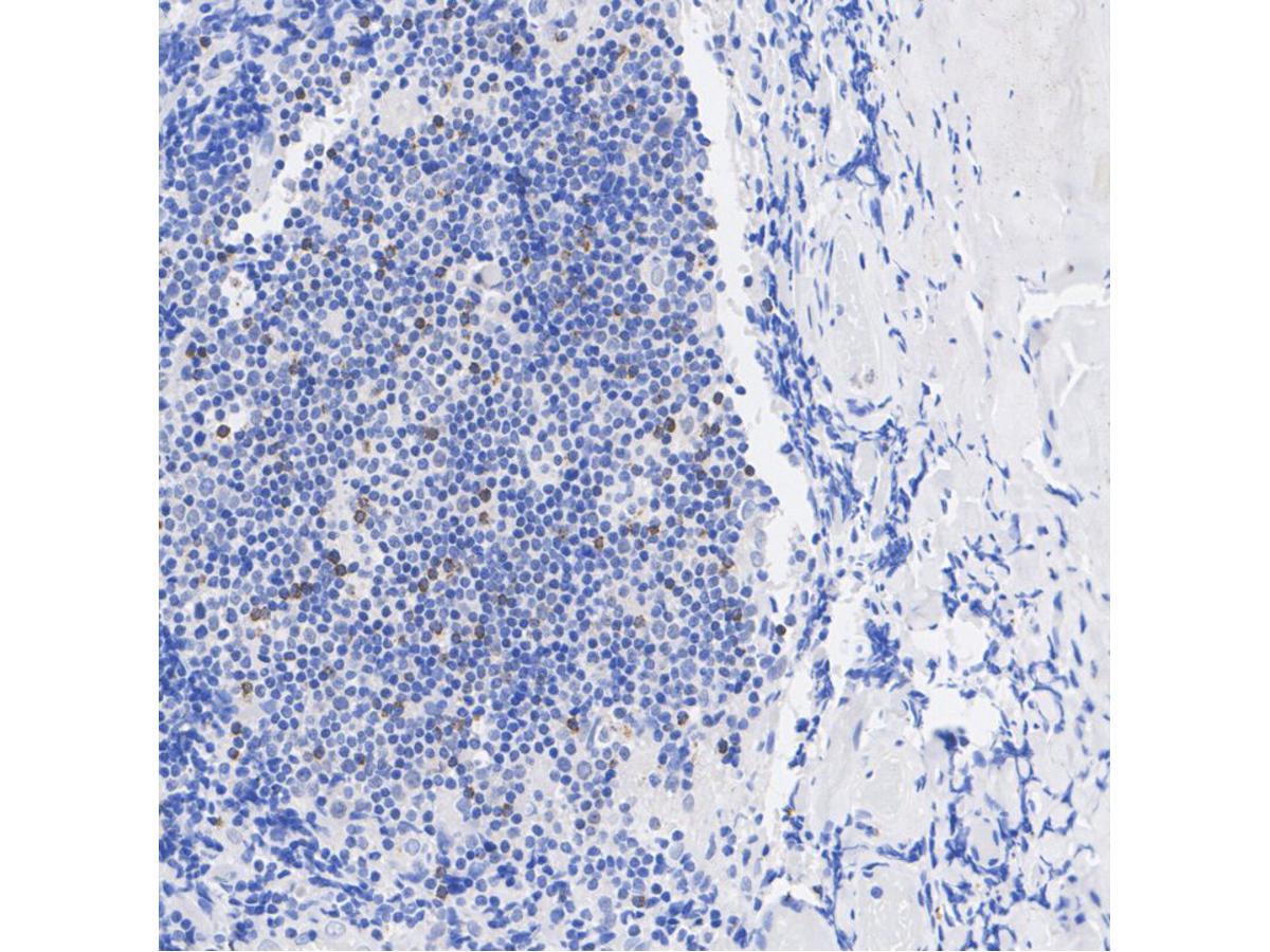
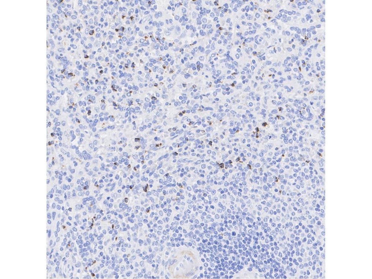
- Immunohistochemical analysis of TIA-1 of paraffin-embedded Human tonsil tissue using a TIA-1 Monoclonal antibody (Product #MA5-32615). Counter stained with hematoxylin.
- Submitted by
- Invitrogen Antibodies (provider)
- Main image
- Experimental details
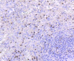
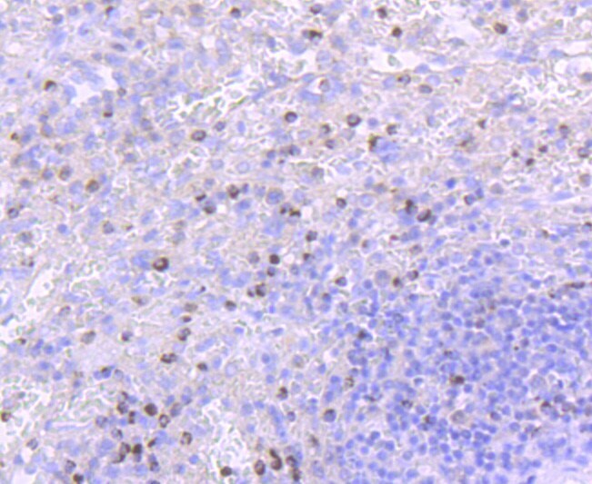
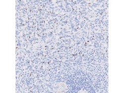
- Immunohistochemical analysis of TIA-1 of paraffin-embedded Human spleen tissue using a TIA-1 Monoclonal antibody (Product #MA5-32615). Counter stained with hematoxylin.
- Submitted by
- Invitrogen Antibodies (provider)
- Main image
- Experimental details
- Immunohistochemistry analysis of paraffin-embedded TIA-1 in human tonsil tissue. The section was pre-treated using heat mediated antigen retrieval with sodium citrate buffer (pH 6.0) for 2 minutes. The tissues were blocked in 1% BSA for 20 minutes at room temperature, washed with ddH2O and PBS. Incubation was done with monoclonal TIA-1 antibody (Product # MA5-32615) at a dilution of 1:200 (1h at RT). The detection was performed using an HRP conjugated compact polymer system. DAB was used as the chromogen. Tissues were counterstained with hematoxylin and mounted with DPX.
- Submitted by
- Invitrogen Antibodies (provider)
- Main image
- Experimental details
- Immunohistochemistry analysis of paraffin-embedded TIA-1 in human spleen tissue. The section was pre-treated using heat mediated antigen retrieval with sodium citrate buffer (pH 6.0) for 2 minutes. The tissues were blocked in 1% BSA for 20 minutes at room temperature, washed with ddH2O and PBS. Incubation was done with monoclonal TIA-1 antibody (Product # MA5-32615) at a dilution of 1:1,000 (1h at RT). The detection was performed using an HRP conjugated compact polymer system. DAB was used as the chromogen. Tissues were counterstained with hematoxylin and mounted with DPX.
Supportive validation
- Submitted by
- Invitrogen Antibodies (provider)
- Main image
- Experimental details
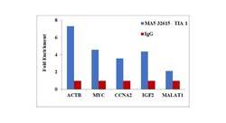
- RNA Immunoprecipitation (RIP) assay of endogenous TIA-1 protein using Anti-TIA-1 Antibody: RIP assay was performed using Anti-TIA-1 Recombinant Rabbit Monoclonal Antibody (Product # MA5-32615, 5 ug) on whole cell lysate from Hep G2 cells exposed to heat shock (45 degrees for 1 hour). Normal Rabbit IgG was used as a negative IP control. RNA purified by RiboPure™ RNA Purification Kit (Product # AM1924) was analyzed by RT-PCR using the Power SYBR® Green RNA-to-CT™ 1-Step Kit (Product # 4389986) with the primers pairs over ACTB, MYC, CCNA2, IGF2 mRNA and MALAT non-coding RNA. Data is presented as fold enrichment of the antibody signal versus the negative control IgG using the comparative CT method.
