Explore
 Validate
 Learn
Learn Western blot
 Immunocytochemistry
Antibody data
- Antibody Data
- Antigen structure
- References [12]
- Comments [0]
- Validations
- Immunocytochemistry [1]
- Immunohistochemistry [1]
Submit
Validation data
Reference
Comment
Report error
- Product number
- HPA007244 - Provider product page

- Provider
- Atlas Antibodies
- Proper citation
- Atlas Antibodies Cat#HPA007244, RRID:AB_1078084
- Product name
- Anti-ACAA1
- Antibody type
- Polyclonal
- Description
- Polyclonal Antibody against Human ACAA1, Gene description: acetyl-CoA acyltransferase 1, Validated applications: WB, ICC, IHC, Uniprot ID: P09110, Storage: Store at +4°C for short term storage. Long time storage is recommended at -20°C.
- Reactivity
- Human, Mouse, Rat
- Host
- Rabbit
- Conjugate
- Unconjugated
- Isotype
- IgG
- Vial size
- 100 µl
- Concentration
- 0.2 mg/ml
- Storage
- Store at +4°C for short term storage. Long time storage is recommended at -20°C.
- Handling
- The antibody solution should be gently mixed before use.
Submitted references Characterization of the Peroxisomal Proteome and Redox Balance in Human Prostate Cancer Cell Lines.
The origin of long-chain fatty acids required for de novo ether lipid/plasmalogen synthesis
Depletion of LONP2 unmasks differential requirements for peroxisomal function between cell types and in cholesterol metabolism
Functional Analysis of GSTK1 in Peroxisomal Redox Homeostasis in HEK-293 Cells.
Autophagy Inhibitors Do Not Restore Peroxisomal Functions in Cells With the Most Common Peroxisome Biogenesis Defect.
Limited survival and impaired hepatic fasting metabolism in mice with constitutive Rag GTPase signaling
HSD17B4, ACAA1, and PXMP4 in Peroxisome Pathway Are Down-Regulated and Have Clinical Significance in Non-small Cell Lung Cancer
Ceapins block the unfolded protein response sensor ATF6α by inducing a neomorphic inter-organelle tether
Peroxisomes contribute to oxidative stress in neurons during doxorubicin-based chemotherapy
Allelic Expression Imbalance Promoting a Mutant PEX6 Allele Causes Zellweger Spectrum Disorder.
Peroxisomal abnormalities in the immortalized human hepatocyte (IHH) cell line.
Peripheral nervous system plasmalogens regulate Schwann cell differentiation and myelination
Hussein MAF, Lismont C, Costa CF, Li H, Claessens F, Fransen M
Antioxidants (Basel, Switzerland) 2024 Nov 1;13(11)
Antioxidants (Basel, Switzerland) 2024 Nov 1;13(11)
The origin of long-chain fatty acids required for de novo ether lipid/plasmalogen synthesis
Chornyi S, Ofman R, Koster J, Waterham H
Journal of Lipid Research 2023;64(5):100364
Journal of Lipid Research 2023;64(5):100364
Depletion of LONP2 unmasks differential requirements for peroxisomal function between cell types and in cholesterol metabolism
Yamashita A, Ignatenko O, Nguyen M, Lambert R, Watt K, Daneault C, Robillard-Frayne I, Topisirovic I, Rosiers C, McBride H
Biology Direct 2023;18(1)
Biology Direct 2023;18(1)
Functional Analysis of GSTK1 in Peroxisomal Redox Homeostasis in HEK-293 Cells.
Costa CF, Lismont C, Chornyi S, Li H, Hussein MAF, Waterham HR, Fransen M
Antioxidants (Basel, Switzerland) 2023 Jun 7;12(6)
Antioxidants (Basel, Switzerland) 2023 Jun 7;12(6)
Autophagy Inhibitors Do Not Restore Peroxisomal Functions in Cells With the Most Common Peroxisome Biogenesis Defect.
Klouwer FCC, Falkenberg KD, Ofman R, Koster J, van Gent D, Ferdinandusse S, Wanders RJA, Waterham HR
Frontiers in cell and developmental biology 2021;9:661298
Frontiers in cell and developmental biology 2021;9:661298
Limited survival and impaired hepatic fasting metabolism in mice with constitutive Rag GTPase signaling
de la Calle Arregui C, Plata-Gómez A, Deleyto-Seldas N, García F, Ortega-Molina A, Abril-Garrido J, Rodriguez E, Nemazanyy I, Tribouillard L, de Martino A, Caleiras E, Campos-Olivas R, Mulero F, Laplante M, Muñoz J, Pende M, Sabio G, Sabatini D, Efeyan A
Nature Communications 2021;12(1)
Nature Communications 2021;12(1)
HSD17B4, ACAA1, and PXMP4 in Peroxisome Pathway Are Down-Regulated and Have Clinical Significance in Non-small Cell Lung Cancer
Zhang X, Yang H, Zhang J, Gao F, Dai L
Frontiers in Genetics 2020;11
Frontiers in Genetics 2020;11
Ceapins block the unfolded protein response sensor ATF6α by inducing a neomorphic inter-organelle tether
Torres S, Gallagher C, Plate L, Gupta M, Liem C, Guo X, Tian R, Stroud R, Kampmann M, Weissman J, Walter P
eLife 2019;8
eLife 2019;8
Peroxisomes contribute to oxidative stress in neurons during doxorubicin-based chemotherapy
Moruno-Manchon J, Uzor N, Kesler S, Wefel J, Townley D, Nagaraja A, Pradeep S, Mangala L, Sood A, Tsvetkov A
Molecular and Cellular Neuroscience 2018;86
Molecular and Cellular Neuroscience 2018;86
Allelic Expression Imbalance Promoting a Mutant PEX6 Allele Causes Zellweger Spectrum Disorder.
Falkenberg KD, Braverman NE, Moser AB, Steinberg SJ, Klouwer FCC, Schlüter A, Ruiz M, Pujol A, Engvall M, Naess K, van Spronsen F, Körver-Keularts I, Rubio-Gozalbo ME, Ferdinandusse S, Wanders RJA, Waterham HR
American journal of human genetics 2017 Dec 7;101(6):965-976
American journal of human genetics 2017 Dec 7;101(6):965-976
Peroxisomal abnormalities in the immortalized human hepatocyte (IHH) cell line.
Klouwer FC, Koster J, Ferdinandusse S, Waterham HR
Histochemistry and cell biology 2017 Apr;147(4):537-541
Histochemistry and cell biology 2017 Apr;147(4):537-541
Peripheral nervous system plasmalogens regulate Schwann cell differentiation and myelination
da Silva T, Eira J, Lopes A, Malheiro A, Sousa V, Luoma A, Avila R, Wanders R, Just W, Kirschner D, Sousa M, Brites P
Journal of Clinical Investigation 2014;124(6):2560-2570
Journal of Clinical Investigation 2014;124(6):2560-2570
No comments: Submit comment
Supportive validation
- Submitted by
- Atlas Antibodies (provider)
- Main image
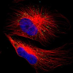
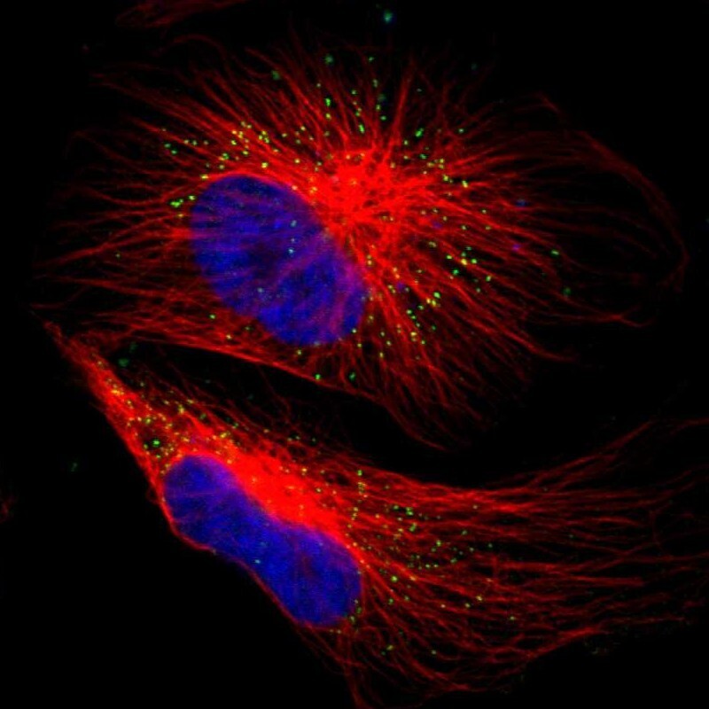
- Experimental details
- Immunofluorescent staining of human cell line U-251 MG shows localization to peroxisomes.
- Sample type
- Human
Supportive validation
- Submitted by
- Atlas Antibodies (provider)
- Enhanced method
- Orthogonal validation
- Main image

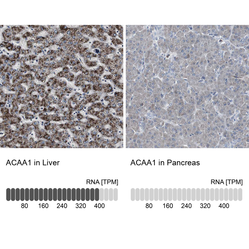
- Experimental details
- Immunohistochemistry analysis in human liver and pancreas tissues using HPA007244 antibody. Corresponding ACAA1 RNA-seq data are presented for the same tissues.
- Sample type
- Human
- Protocol
- Protocol