 Explore
 Validate
 Learn
Learn Western blot
Antibody data
- Antibody Data
- Antigen structure
- References [1]
- Comments [0]
- Validations
- Western blot [3]
- Immunocytochemistry [1]
Submit
Validation data
Reference
Comment
Report error
- Product number
- PA1-41122 - Provider product page

- Provider
- Invitrogen Antibodies
- Product name
- HTR7 Polyclonal Antibody
- Antibody type
- Polyclonal
- Antigen
- Synthetic peptide
- Description
- This antibody is predicted to react with porcine samples based on sequence homology.
- Concentration
- 1 mg/mL
Submitted references Creation of the 5-hydroxytryptamine receptor 7 knockout rat as a tool for cardiovascular research.
Demireva EY, Xie H, Flood ED, Thompson JM, Seitz BM, Watts SW
Physiological genomics 2019 Jul 1;51(7):290-301
Physiological genomics 2019 Jul 1;51(7):290-301
No comments: Submit comment
Supportive validation
- Submitted by
- Invitrogen Antibodies (provider)
- Main image
- Experimental details
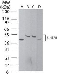
- Western blot analysis of 5-HT7R in A) human brain, B) mouse brain, C) rat brain, and D) humanSK-N-SHneuroblastoma cell lysate using a Serotonin Receptor 7 polyclonal antibody (Product # PA1-41122) at 2 µg/mL.

- Submitted by
- Invitrogen Antibodies (provider)
- Main image

- Experimental details
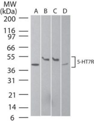
- Western blot analysis of HTR7 in A) human brain, B) mouse brain, C) rat brain, and D) human SK-N-SH neuroblastoma cell lysate. Samples were incubated in HTR7 polyclonal antibody (Product # PA1-41122).
- Submitted by
- Invitrogen Antibodies (provider)
- Main image
- Experimental details
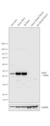
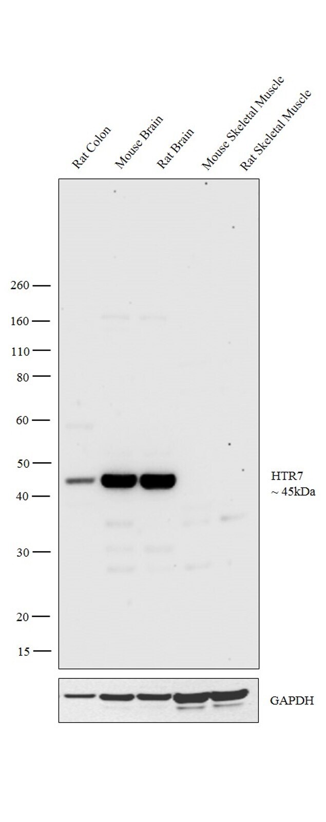
- Western blot analysis was performed on tissue extracts (30 µg lysate) of Rat Colon (Lane 1), Mouse Brain (Lane 2), Rat Brain (Lane 3), Mouse Skeletal Muscle (Lane 4) and Rat Skeletal Muscle Raji (Lane 5). The blot was probed with Anti-HTR7 Polyclonal Antibody (Product # PA1-41122, 1µg/ml) and detected by chemiluminescence using Goat anti-Rabbit IgG (H+L) Superclonal™ Secondary Antibody, HRP conjugate (Product # A27036, 0.25 µg/ml, 1:4000 dilution). A 45kDa band corresponding to HTR7 was observed across all tissue extracts tested except for Mouse Skeletal Muscle and Rat Skeletal Muscle.
Supportive validation
- Submitted by
- Invitrogen Antibodies (provider)
- Main image
- Experimental details
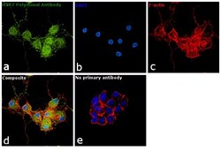
- Immunofluorescence analysis of HTR7 was performed using 70% confluent log phase differentiated PC-12 cells into neurons. The cells were fixed with 4% paraformaldehyde for 10 minutes, permeabilized with 0.1% Triton™ X-100 for 15 minutes, and blocked with 1% BSA for 1 hour at room temperature. The cells were labeled with HTR7 Polyclonal Antibody (Product # PA1-41122) at 1:100 dilution in 0.1% BSA, incubated at 4 degree Celsius overnight and then labeled with Goat anti-Rabbit IgG (H+L) Superclonal™ Secondary Antibody, Alexa Fluor® 488 conjugate (Product # A27034) at a dilution of 1:2000 for 45 minutes at room temperature (Panel a: green). Nuclei (Panel b: blue) were stained with ProLong™ Diamond Antifade Mountant with DAPI (Product # P36962). F-actin (Panel c: red) was stained with Rhodamine Phalloidin (Product # R415). Panel d represents the merged image showing cytoplasmic and membrane localization. Panel e represents control cells with no primary antibody to assess background. The images were captured at 60X magnification.
