Explore
 Validate
 Learn
Learn Western blot
 Immunocytochemistry
Antibody data
- Antibody Data
- Antigen structure
- References [1]
- Comments [0]
- Validations
- Western blot [7]
- Immunocytochemistry [1]
- Immunoprecipitation [1]
- Immunohistochemistry [1]
Submit
Validation data
Reference
Comment
Report error
- Product number
- GTX104122 - Provider product page

- Provider
- GeneTex
- Proper citation
- GeneTex Cat#GTX104122, RRID:AB_1950905
- Product name
- ME1 antibody
- Antibody type
- Polyclonal
- Reactivity
- Human, Mouse, Rat
- Host
- Rabbit
Submitted references A clinical drug library screen identifies clobetasol propionate as an NRF2 inhibitor with potential therapeutic efficacy in KEAP1 mutant lung cancer.
Choi EJ, Jung BJ, Lee SH, Yoo HS, Shin EA, Ko HJ, Chang S, Kim SY, Jeon SM
Oncogene 2017 Sep 14;36(37):5285-5295
Oncogene 2017 Sep 14;36(37):5285-5295
No comments: Submit comment
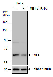
Enhanced validation
Supportive validation
- Submitted by
- GeneTex (provider)
- Enhanced method
- Genetic validation
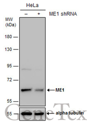
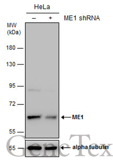
- Main image
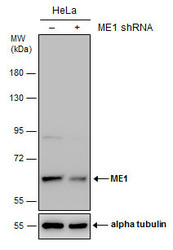
- Experimental details
- Non-transfected (¡V) and transfected (+) HeLa whole cell extracts (30 ?g) were separated by 7.5% SDS-PAGE, and the membrane was blotted with ME1 antibody (GTX104122) diluted at 1:6000. The HRP-conjugated anti-rabbit IgG antibody (GTX213110-01) was used to detect the primary antibody.
Supportive validation
- Submitted by
- GeneTex (provider)
- Main image
- Experimental details
- Sample (30 ?g of whole cell lysate) A: PC-12 7.5% SDS PAGE GTX104122 diluted at 1:1000 The HRP-conjugated anti-rabbit IgG antibody (GTX213110-01) was used to detect the primary antibody.
- Submitted by
- GeneTex (provider)
- Main image
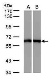
- Experimental details
- Sample(30 ug whole cell lysate)A: A431(GTX27909)B: HeLa S3(GTX14654)7.5% SDS PAGEGTX104122 diluted at 1:500
- Validation comment
- WB
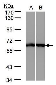
- Submitted by
- GeneTex (provider)
- Main image
- Experimental details
- Sample (30 ?g of whole cell lysate) A: NIH-3T3 B: JC C: BCL-1 7.5% SDS PAGE GTX104122 diluted at 1:1000 The HRP-conjugated anti-rabbit IgG antibody (GTX213110-01) was used to detect the primary antibody.

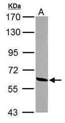
- Submitted by
- GeneTex (provider)
- Main image
- Experimental details
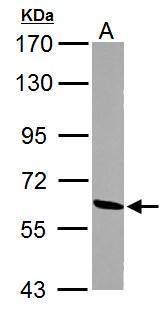
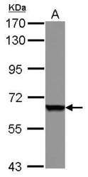
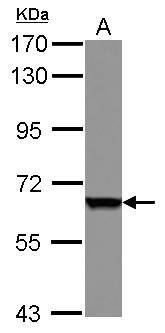
- Sample (30 ug of whole cell lysate) A: A431 7.5% SDS PAGE GTX104122 diluted at 1:1000
- Validation comment
- WB
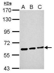
- Submitted by
- GeneTex (provider)
- Main image
- Experimental details
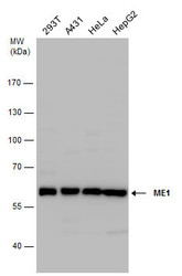
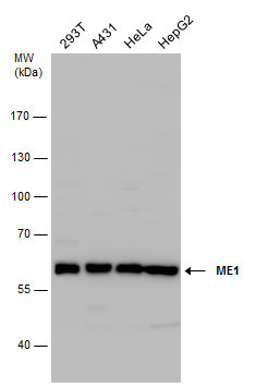
- ME1 antibody detects ME1 protein by western blot analysis. Various whole cell extracts (30 ?g) were separated by 7.5% SDS-PAGE, and the membrane was blotted with ME1 antibody (GTX104122) diluted at a dilution of 1:1000. The HRP-conjugated anti-rabbit IgG antibody (GTX213110-01) was used to detect the primary antibody.
- Submitted by
- GeneTex (provider)
- Main image
- Experimental details
- Non-transfected (¡V) and transfected (+) HeLa whole cell extracts (30 ?g) were separated by 7.5% SDS-PAGE, and the membrane was blotted with ME1 antibody (GTX104122) diluted at 1:6000. The HRP-conjugated anti-rabbit IgG antibody (GTX213110-01) was used to detect the primary antibody.
Supportive validation
- Submitted by
- GeneTex (provider)
- Main image
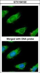
- Experimental details
- Immunofluorescence analysis of paraformaldehyde-fixed HeLa, using ME1(GTX104122) antibody at 1:200 dilution.

Supportive validation
- Submitted by
- GeneTex (provider)
- Main image
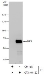
- Experimental details
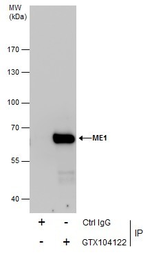
- Immunoprecipitation of ME1 protein from HeLa whole cell extracts using 5 £gg of ME1 antibody (GTX104122).Western blot analysis was performed using ME1 antibody (GTX104122).EasyBlot anti-Rabbit IgG (GTX221666-01) was used as a secondary reagent.
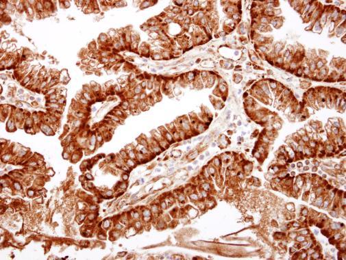
Supportive validation
- Submitted by
- GeneTex (provider)
- Main image

- Experimental details
- ME1 antibody detects ME1 protein at cytosol on human ovarian carcinoma by immunohistochemical analysis. Sample: Paraffin-embedded human ovarian carcinoma. ME1 antibody (GTX104122) dilution: 1:500.