Explore
 Validate
 Learn
Learn Western blot
 Immunocytochemistry
Antibody data
- Antibody Data
- Antigen structure
- References [1]
- Comments [0]
- Validations
- Immunocytochemistry [1]
- Immunohistochemistry [1]
Submit
Validation data
Reference
Comment
Report error
- Product number
- HPA008247 - Provider product page

- Provider
- Atlas Antibodies
- Proper citation
- Atlas Antibodies Cat#HPA008247, RRID:AB_1853704
- Product name
- Anti-ME2
- Antibody type
- Polyclonal
- Description
- Polyclonal Antibody against Human ME2, Gene description: malic enzyme 2, NAD(+)-dependent, mitochondrial, Validated applications: WB, IHC, ICC, Uniprot ID: P23368, Storage: Store at +4°C for short term storage. Long time storage is recommended at -20°C.
- Reactivity
- Human
- Host
- Rabbit
- Conjugate
- Unconjugated
- Isotype
- IgG
- Vial size
- 100 µl
- Concentration
- 0.1 mg/ml
- Storage
- Store at +4°C for short term storage. Long time storage is recommended at -20°C.
- Handling
- The antibody solution should be gently mixed before use.
Submitted references Genomic deletion of malic enzyme 2 confers collateral lethality in pancreatic cancer
Dey P, Baddour J, Muller F, Wu C, Wang H, Liao W, Lan Z, Chen A, Gutschner T, Kang Y, Fleming J, Satani N, Zhao D, Achreja A, Yang L, Lee J, Chang E, Genovese G, Viale A, Ying H, Draetta G, Maitra A, Wang Y, Nagrath D, DePinho R
Nature 2017;542(7639):119-123
Nature 2017;542(7639):119-123
No comments: Submit comment
Supportive validation
- Submitted by
- Atlas Antibodies (provider)
- Main image

- Experimental details
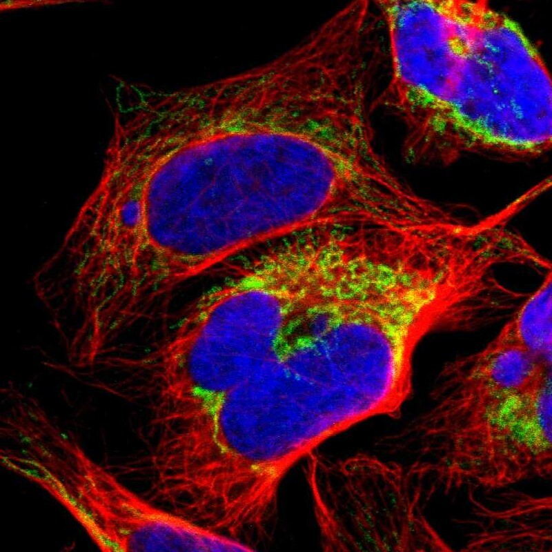
- Immunofluorescent staining of human cell line U-2 OS shows localization to mitochondria.
- Sample type
- Human
Supportive validation
- Submitted by
- Atlas Antibodies (provider)
- Enhanced method
- Orthogonal validation
- Main image
- Experimental details
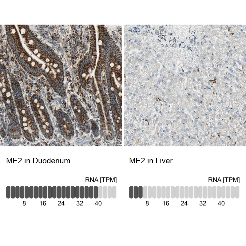
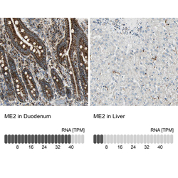
- Immunohistochemistry analysis in human duodenum and liver tissues using Anti-ME2 antibody. Corresponding ME2 RNA-seq data are presented for the same tissues.
- Sample type
- Human
- Protocol
- Protocol