Explore
 Validate
 Learn
Learn Western blot
 Immunocytochemistry
Antibody data
- Antibody Data
- Antigen structure
- References [2]
- Comments [0]
- Validations
- Immunocytochemistry [3]
- Immunohistochemistry [1]
- Other assay [1]
Submit
Validation data
Reference
Comment
Report error
- Product number
- PA5-20272 - Provider product page

- Provider
- Invitrogen Antibodies
- Product name
- TIP47 Polyclonal Antibody
- Antibody type
- Polyclonal
- Antigen
- Synthetic peptide
- Description
- A suggested positive control is Daudi cell lysate. PA5-20272 can be used with blocking peptide PEP-0387.
- Reactivity
- Human, Rat
- Host
- Rabbit
- Isotype
- IgG
- Vial size
- 100 μg
- Concentration
- 1 mg/mL
- Storage
- Maintain refrigerated at 2-8°C for up to 3 months. For long term storage store at -20°C
Submitted references Real-Time Tracking of BODIPY-C12 Long-Chain Fatty Acid in Human Term Placenta Reveals Unique Lipid Dynamics in Cytotrophoblast Cells.
Myotubes from severely obese type 2 diabetic subjects accumulate less lipids and show higher lipolytic rate than myotubes from severely obese non-diabetic subjects.
Kolahi K, Louey S, Varlamov O, Thornburg K
PloS one 2016;11(4):e0153522
PloS one 2016;11(4):e0153522
Myotubes from severely obese type 2 diabetic subjects accumulate less lipids and show higher lipolytic rate than myotubes from severely obese non-diabetic subjects.
Bakke SS, Feng YZ, Nikolić N, Kase ET, Moro C, Stensrud C, Damlien L, Ludahl MO, Sandbu R, Solheim BM, Rustan AC, Hjelmesæth J, Thoresen GH, Aas V
PloS one 2015;10(3):e0119556
PloS one 2015;10(3):e0119556
No comments: Submit comment
Supportive validation
- Submitted by
- Invitrogen Antibodies (provider)
- Main image
- Experimental details
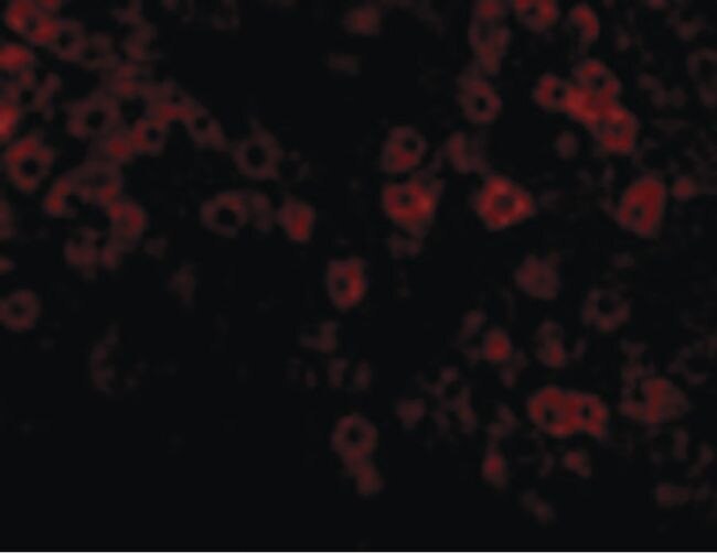
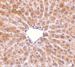
- Immunofluorescent analysis of rat liver cells using a TIP47 polyclonal antibody (Product # PA5-20272) at a 20 µg/mL dilution.
- Submitted by
- Invitrogen Antibodies (provider)
- Main image
- Experimental details
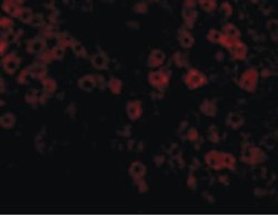
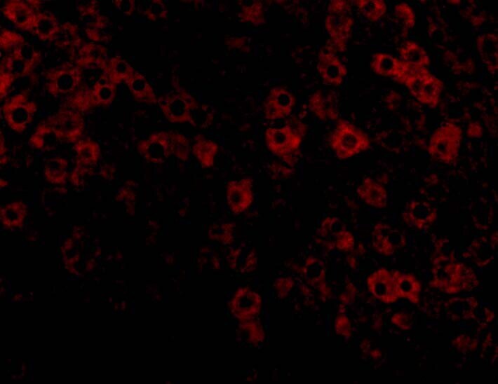
- Immunofluorescent analysis of 4% paraformaldehyde-fixed rat liver cells labeling TIP47 with TIP47 Polyclonal Antibody (Product # PA5-20272) at 20 µg/mL, followed by goat anti-rabbit IgG secondary antibody at 1:500 dilution (red).
- Submitted by
- Invitrogen Antibodies (provider)
- Main image

- Experimental details
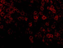
- Immunofluorescent analysis of 4% paraformaldehyde-fixed rat liver cells labeling TIP47 with TIP47 Polyclonal Antibody (Product # PA5-20272) at 20 µg/mL, followed by goat anti-rabbit IgG secondary antibody at 1:500 dilution (red).

Supportive validation
- Submitted by
- Invitrogen Antibodies (provider)
- Main image
- Experimental details
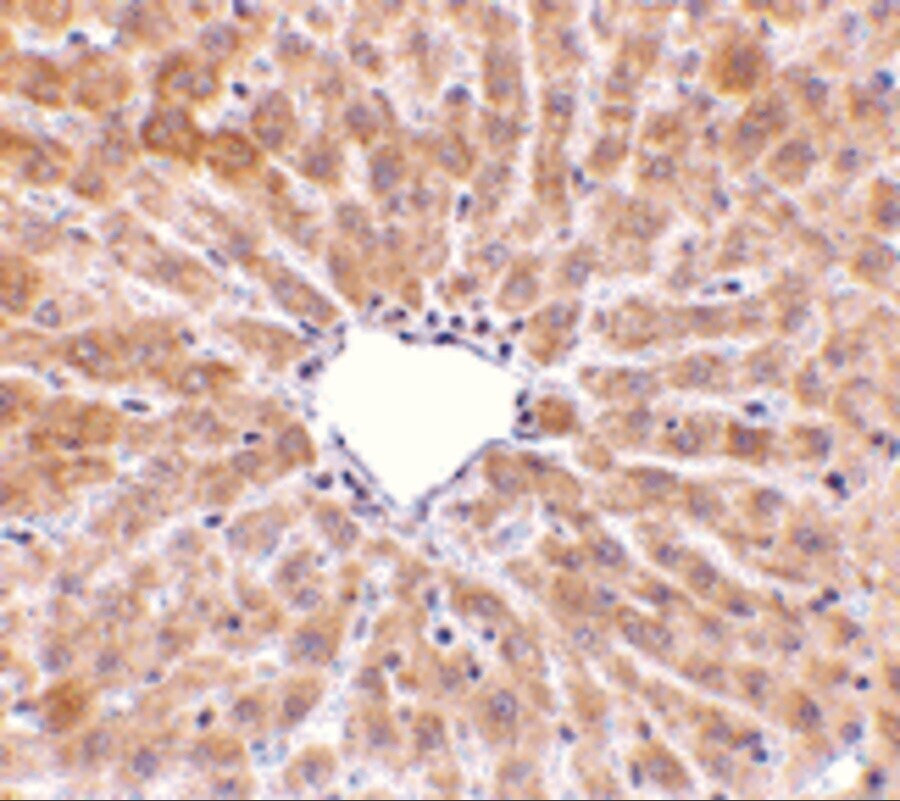
- Immunohistochemical analysis of paraffin-embedded Rat Liver Tissue using TIP47 Polyclonal Antibody (Product # PA5-20272) at 10 µg/mL. Tissue was fixed with formaldehyde and blocked with 0.1 serum for 1 h at RT; antigen retrieval was by heat mediation with a citrate buffer (pH6). Samples were incubated with primary antibody overnight at 4˚C. A goat anti-rabbit IgG H&L (HRP) at 1/250 was used as secondary. Counter stained with Hematoxylin.
Supportive validation
- Submitted by
- Invitrogen Antibodies (provider)
- Main image

- Experimental details
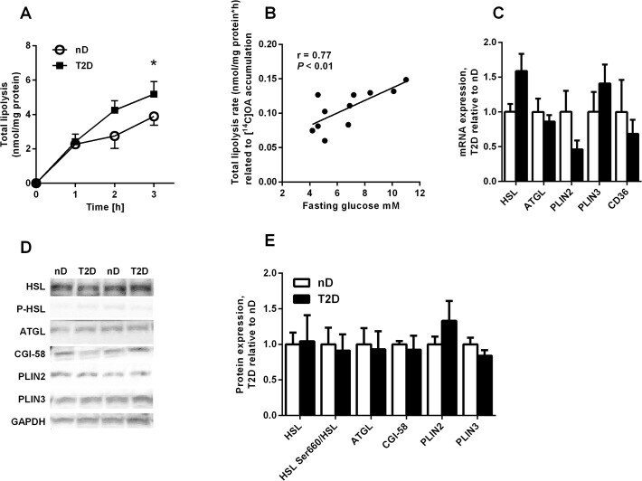
- Fig 3 Higher lipolysis rate in myotubes from severely obese donors with type 2 diabetes. ( A ) Total lipolysis (with triacsin C) after 24 h incubation with [ 14 C]OA in non-diabetic (nD) and type 2 diabetic myotubes (T2D) n = 5-6. ( B ) Fasting plasma glucose levels correlated positively with total lipolysis rate, n = 11 (C) mRNA expression of hormone-sensitive lipase (HSL), adipose triglyceride lipase (ATGL), perilipin 2 (PLIN2), perilipin 3 (PLIN3) and the fatty acid transporter CD36. Data are presented relative to mean values of non-diabetics, n = 6-10. ( D-E ) Protein expression of HSL, ATGL, comparative gene identification 58 (CGI-58), PLIN2 and PLIN3 and phosphorylation of HSL at serine 660 (HSL Ser660) after 24h incubation with 100 muM OA. (D) Two representative blots are shown. Data are presented relative to mean values of non-diabetics, n = 5.