Explore
 Validate
 Learn
Learn Western blot
Antibody data
- Antibody Data
- Antigen structure
- References [2]
- Comments [0]
- Validations
- Western blot [2]
- Immunohistochemistry [1]
- Other assay [1]
Submit
Validation data
Reference
Comment
Report error
- Product number
- PA5-69347 - Provider product page

- Provider
- Invitrogen Antibodies
- Product name
- CPT1A Polyclonal Antibody
- Antibody type
- Polyclonal
- Antigen
- Synthetic peptide
- Description
- This target displays homology in the following species: Cow: 100%; Dog: 100%; Guinea Pig: 100%; Horse: 100%; Human: 100%; Mouse: 100%; Rabbit: 100%; Rat: 100%; Sheep: 100%; Zebrafish: 100%
- Reactivity
- Human
- Host
- Rabbit
- Isotype
- IgG
- Vial size
- 100 µL
- Concentration
- 0.5 mg/mL
- Storage
- -20° C, Avoid Freeze/Thaw Cycles
Submitted references Fatty acid β-oxidation promotes breast cancer stemness and metastasis via the miRNA-328-3p-CPT1A pathway.
Downregulating carnitine palmitoyl transferase 1 affects disease progression in the SOD1 G93A mouse model of ALS.
Zeng F, Yao M, Wang Y, Zheng W, Liu S, Hou Z, Cheng X, Sun S, Li T, Zhao H, Luo Y, Li J
Cancer gene therapy 2022 Mar;29(3-4):383-395
Cancer gene therapy 2022 Mar;29(3-4):383-395
Downregulating carnitine palmitoyl transferase 1 affects disease progression in the SOD1 G93A mouse model of ALS.
Trabjerg MS, Andersen DC, Huntjens P, Oklinski KE, Bolther L, Hald JL, Baisgaard AE, Mørk K, Warming N, Kullab UB, Kroese LJ, Pritchard CEJ, Huijbers IJ, Nieland JDV
Communications biology 2021 Apr 30;4(1):509
Communications biology 2021 Apr 30;4(1):509
No comments: Submit comment
Supportive validation
- Submitted by
- Invitrogen Antibodies (provider)
- Main image
- Experimental details
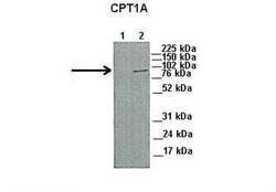
- Western blot analysis of CPT1A on capan1 and HPAF cells using 45 µg of sample per lane (Lane 1: capan1 cell lysate, 2: HPAF cell lysate). The sample was probed with a CPT1A polyclonal antibody (Product # PA5-69347) using a primary antibody dilution of 1:1000 and a secondary antibody dilution of 1:5000.
- Submitted by
- Invitrogen Antibodies (provider)
- Main image
- Experimental details
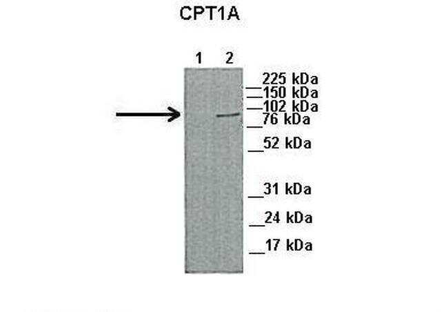
- Western blot analysis of CPT1A on human 293T cells. The sample was probed with a CPT1A polyclonal antibody (Product # PA5-69347) using a primary antibody dilution of 0.2-1.0 µg/mL.

Supportive validation
- Submitted by
- Invitrogen Antibodies (provider)
- Main image
- Experimental details
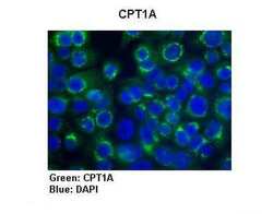
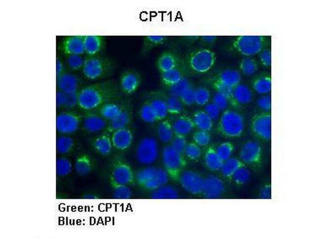
- Immunohistochemistry was performed on human pancreatic cells tissue using a CPT1A polyclonal primary antibody (Product # PA5-69347) with a dilution of 1:300, anti-rabbit Alexa Fluor 488 with a secondary antibody dilution of 1:200 and CPT1A (green), and DAPI (blue).
Supportive validation
- Submitted by
- Invitrogen Antibodies (provider)
- Main image
- Experimental details
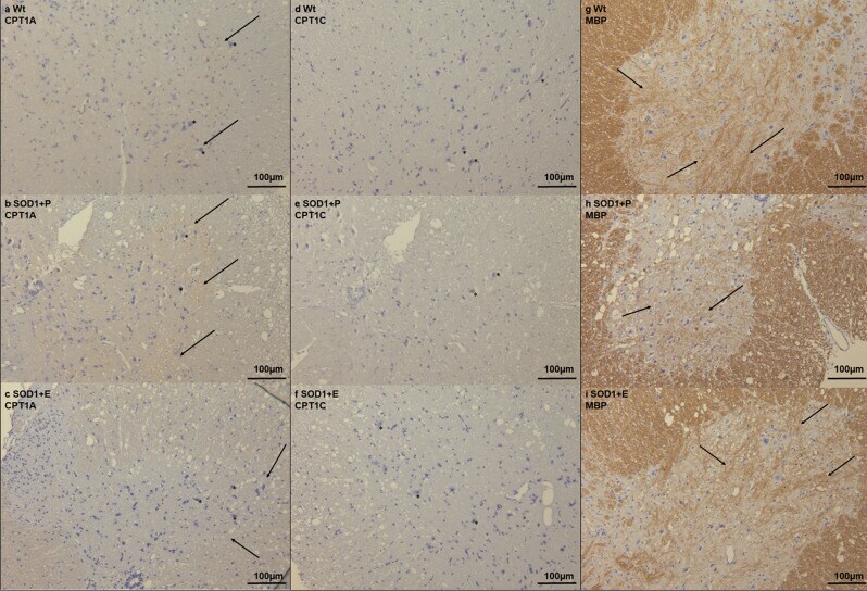
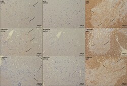
- Fig. 3 Immunohistochemical staining in the lumbar spinal from the SOD1 G93A etomoxir experiment. a - c CPT1A staining in lumbar spinal cord from Wt, SOD1 + P and SOD1 + E mice at day 130 indicating increased labeling in SOD1 + P mice (arrows) and pathological morphology of neurons (asterisks). d - f CPT1C staining in lumbar spinal cord from Wt, SOD1 + P and SOD1 + E mice at day 130 indicating no difference in the labeling in SOD1 + P mice but differences in the morphology of neurons (asterisks). g - i MBP staining in lumbar spinal cord from Wt, SOD1 + P and SOD1 + E mice at day 130 indicating decreased labeling in SOD1 + P mice (arrows). All images are presented with 16x magnification. N = 2-4 animals per group. WT = wild-type, SOD1 = SOD1 G93A genotype, E = etomoxir, P = placebo.