 Explore
 Validate
 Learn
Learn Western blot
 Immunohistochemistry
Antibody data
- Antibody Data
- Antigen structure
- References [4]
- Comments [0]
- Validations
- Immunohistochemistry [1]
Submit
Validation data
Reference
Comment
Report error
- Product number
- HPA011990 - Provider product page

- Provider
- Atlas Antibodies
- Proper citation
- Atlas Antibodies Cat#HPA011990, RRID:AB_1844465
- Product name
- Anti-ACADL
- Antibody type
- Polyclonal
- Description
- Polyclonal Antibody against Human ACADL, Gene description: acyl-CoA dehydrogenase, long chain, Alternative Gene Names: ACAD4, LCAD, Validated applications: IHC, WB, Uniprot ID: P28330, Storage: Store at +4°C for short term storage. Long time storage is recommended at -20°C.
- Reactivity
- Human, Mouse, Rat
- Host
- Rabbit
- Conjugate
- Unconjugated
- Isotype
- IgG
- Vial size
- 100 µl
- Concentration
- 0.1 mg/ml
- Storage
- Store at +4°C for short term storage. Long time storage is recommended at -20°C.
- Handling
- The antibody solution should be gently mixed before use.
Submitted references ACADL-YAP axis activity in non-small cell lung cancer carcinogenicity
Meningioma classification by immunohistochemistry: A replicability study.
A clinically applicable integrative molecular classification of meningiomas
ACADL plays a tumor-suppressor role by targeting Hippo/YAP signaling in hepatocellular carcinoma
Chen K, Hong C, Kong W, Li G, Liu Z, Zhu K, Lu C, Si P, Gao P, Ning G, Zhang R
Cancer Cell International 2024;24(1)
Cancer Cell International 2024;24(1)
Meningioma classification by immunohistochemistry: A replicability study.
Näslund O, Lipatnikova A, Dénes A, Lindskog C, Bontell TO, Smits A, Jakola AS, Corell A
Brain & spine 2023;3:101711
Brain & spine 2023;3:101711
A clinically applicable integrative molecular classification of meningiomas
Nassiri F, Liu J, Patil V, Mamatjan Y, Wang J, Hugh-White R, Macklin A, Khan S, Singh O, Karimi S, Corona R, Liu L, Chen C, Chakravarthy A, Wei Q, Mehani B, Suppiah S, Gao A, Workewych A, Tabatabai G, Boutros P, Bader G, de Carvalho D, Kislinger T, Aldape K, Zadeh G
Nature 2021;597(7874):119-125
Nature 2021;597(7874):119-125
ACADL plays a tumor-suppressor role by targeting Hippo/YAP signaling in hepatocellular carcinoma
Zhao X, Qin W, Jiang Y, Yang Z, Yuan B, Dai R, Shen H, Chen Y, Fu J, Wang H
npj Precision Oncology 2020;4(1)
npj Precision Oncology 2020;4(1)
No comments: Submit comment
Supportive validation
- Submitted by
- Atlas Antibodies (provider)
- Enhanced method
- Orthogonal validation
- Main image
- Experimental details
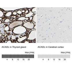
- Immunohistochemistry analysis in human thyroid gland and cerebral cortex tissues using HPA011990 antibody. Corresponding ACADL RNA-seq data are presented for the same tissues.
- Sample type
- Human
- Protocol
- Protocol
