Explore
 Validate
 Learn
LearnPA5-27480
antibody from Invitrogen Antibodies
Targeting: GRK2
ADRBK1, BARK1
 Western blot Immunocytochemistry
 Immunoprecipitation Immunohistochemistry
 Flow cytometry Other assay
Antibody data
- Antibody Data
- Antigen structure
- References [1]
- Comments [0]
- Validations
- Western blot [6]
- Immunocytochemistry [2]
- Immunohistochemistry [1]
- Other assay [2]
Submit
Validation data
Reference
Comment
Report error
- Product number
- PA5-27480 - Provider product page

- Provider
- Invitrogen Antibodies
- Product name
- GRK2 Polyclonal Antibody
- Antibody type
- Polyclonal
- Antigen
- Recombinant protein fragment
- Description
- Recommended positive controls: 293T, A431, HeLaS3, HepG2, Molt-4, Raji, NIH-3T3, Jurkat, NCI-H929. Predicted reactivity: Mouse (98%), Rat (98%), Pig (98%), Bovine (98%). Store product as a concentrated solution. Centrifuge briefly prior to opening the vial.
- Reactivity
- Human, Mouse
- Host
- Rabbit
- Isotype
- IgG
- Vial size
- 100 µL
- Concentration
- 0.82 mg/mL
- Storage
- Store at 4°C short term. For long term storage, store at -20°C, avoiding freeze/thaw cycles.
Submitted references Alterations of the renin angiotensin system in human end-stage heart failure before and after mechanical cardiac unloading by LVAD support.
Messmann R, Dietl A, Wagner S, Domenig O, Jungbauer C, Luchner A, Maier LS, Schopka S, Hirt S, Schmid C, Birner C
Molecular and cellular biochemistry 2020 Sep;472(1-2):79-94
Molecular and cellular biochemistry 2020 Sep;472(1-2):79-94
No comments: Submit comment
Supportive validation
- Submitted by
- Invitrogen Antibodies (provider)
- Main image
- Experimental details
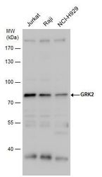
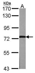
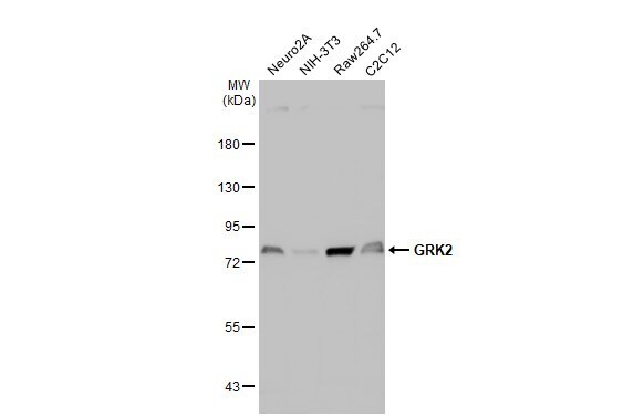
- Western blot analysis of GRK2 using Various whole cell extracts (30 µg). Samples were loaded onto a 7.5% SDS-PAGE gel and probed with a GRK2 polyclonal antibody (Product # PA5-27480) at a dilution of 1:1000.

- Submitted by
- Invitrogen Antibodies (provider)
- Main image
- Experimental details
- Western blot analysis of GRK2 using 30 µg of MOLT4 lysate. Samples were loaded onto a 7.5% SDS-PAGE gel and probed with a GRK2 polyclonal antibody (Product # PA5-27480) at a dilution of 1:1000.
- Submitted by
- Invitrogen Antibodies (provider)
- Main image
- Experimental details
- Western blot analysis of GRK2 using 30 µg of NIH-3T3 lysate. Samples were loaded onto a 7.5% SDS-PAGE gel and probed with a GRK2 polyclonal antibody (Product # PA5-27480) at a dilution of 1:1000.

- Submitted by
- Invitrogen Antibodies (provider)
- Main image
- Experimental details
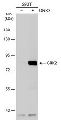
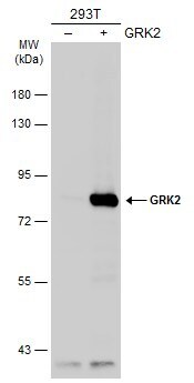
- Western Blot analysis of GRK2 was performed by separating 30 µg of non-transfected (–) and transfected (+) 293T whole cell extracts by 5% SDS-PAGE. Proteins were transferred to a membrane and probed with a GRK2 Polyclonal Antibody (Product # PA5-27480) at a dilution of 1:1000. The HRP-conjugated anti-rabbit IgG antibody was used to detect the primary antibody.
- Submitted by
- Invitrogen Antibodies (provider)
- Main image
- Experimental details
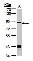
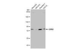
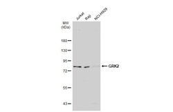
- Western Blot using GRK2 Polyclonal Antibody (Product # PA5-27480). Various whole cell extracts (30 µg) were separated by 12% SDS-PAGE, and the membrane was blotted with GRK2 Polyclonal Antibody (Product # PA5-27480) diluted at 1:1,000. The HRP-conjugated anti-rabbit IgG antibody was used to detect the primary antibody.
- Submitted by
- Invitrogen Antibodies (provider)
- Main image
- Experimental details
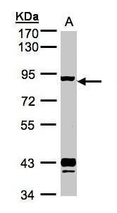
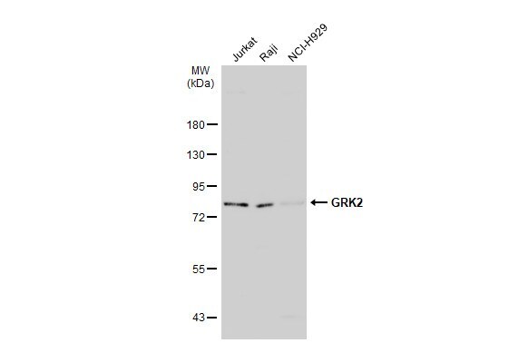
- Western Blot using GRK2 Polyclonal Antibody (Product # PA5-27480). Various whole cell extracts (30 µg) were separated by 7.5% SDS-PAGE, and the membrane was blotted with GRK2 Polyclonal Antibody (Product # PA5-27480) diluted at 1:1,000. The HRP-conjugated anti-rabbit IgG antibody was used to detect the primary antibody.
Supportive validation
- Submitted by
- Invitrogen Antibodies (provider)
- Main image

- Experimental details
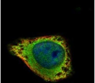
- Immunofluorescent analysis of GRK2 in methanol-fixed HeLa cells using a GRK2 polyclonal antibody (Product # PA5-27480) (Green) at a 1:100 dilution. Alpha-tubulin filaments were labeled with Product # PA5-29281 (Red) at a 1:2000.
- Submitted by
- Invitrogen Antibodies (provider)
- Main image

- Experimental details
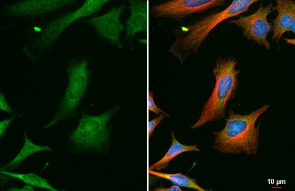
- GRK2 Polyclonal Antibody detects GRK2 protein at cytoplasm by immunofluorescent analysis. Sample: HeLa cells were fixed in ice-cold MeOH for 5 min. Green: GRK2 stained by GRK2 Polyclonal Antibody (Product # PA5-27480) diluted at 1:1,000. Red: alpha Tubulin, a cytoskeleton marker, stained by alpha Tubulin Polyclonal Antibody [GT114] (Product # MA5-31466) diluted at 1:1,000. Blue: Fluoroshield with DAPI .
Supportive validation
- Submitted by
- Invitrogen Antibodies (provider)
- Main image
- Experimental details
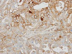
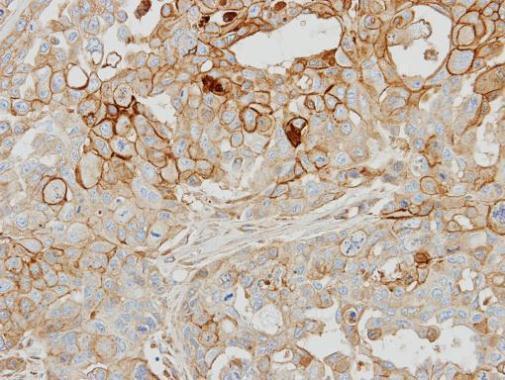
- Immunohistochemical analysis of paraffin-embedded OVCAR3 xenograft, using GRK2 (Product # PA5-27480) antibody at 1:500 dilution. Antigen Retrieval: Citrate buffer, pH 6.0, 15 min.
Supportive validation
- Submitted by
- Invitrogen Antibodies (provider)
- Main image
- Experimental details
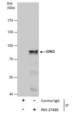
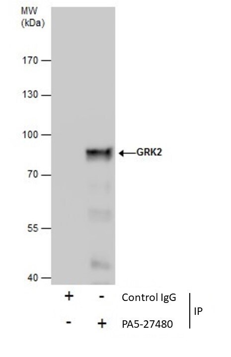
- Immunoprecipitation of GRK2 was performed in Jurkat whole cell extracts using 5 µg of GRK2 Polyclonal Antibody (Product # PA5-27480). Samples were transferred to a membrane and probed with GRK2 Polyclonal Antibody as a primary antibody and an HRP-conjugated anti-Rabbit IgG was used as a secondary antibody.
- Submitted by
- Invitrogen Antibodies (provider)
- Main image
- Experimental details
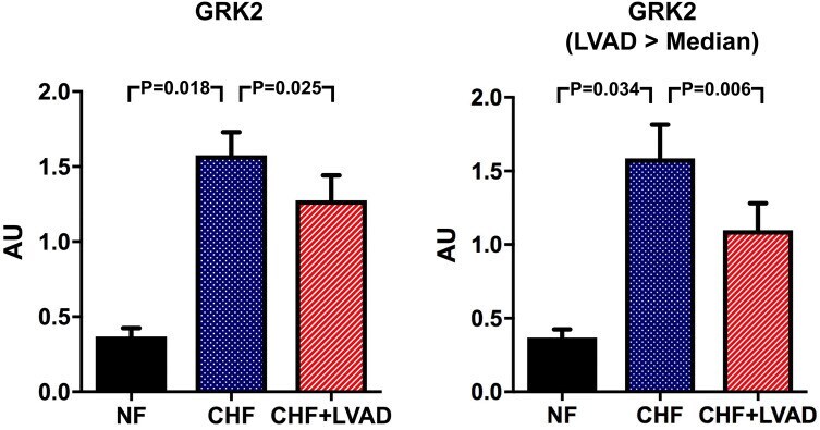
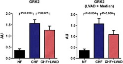
- Fig. 7 GRK2 before (CHF) and after LVAD therapy (CHF+LVAD) as compared to non-failing ventricles (NF), left. The same analysis for the subgroups of patients with a duration of LVAD therapy above the median value, right ( n = 10). GRK2 expression was determined by immunoblot (western blot) analysis and referred to a standard, respectively, whose densitometric value was set 1 by default. The value on the y -axis, therefore, reflects the percentage of each parameter's immunoblot band density in relation to this default value. AU arbitrary unit, CHF congestive heart failure, LVAD left ventricular assist device, NF non-failing myocardial tissue specimen