Explore
 Validate
 Learn
Learn Western blot
Antibody data
- Antibody Data
- Antigen structure
- References [2]
- Comments [0]
- Validations
- Western blot [3]
- Immunocytochemistry [1]
- Immunohistochemistry [1]
Submit
Validation data
Reference
Comment
Report error
- Product number
- AP8093b - Provider product page

- Provider
- Abcepta
- Proper citation
- Abgent Cat#AP8093b, RRID:AB_2160003
- Product name
- PCK1 Antibody (C-term)
- Antibody type
- Polyclonal
- Antigen
- Synthetic peptide
- Description
- Purified Rabbit Polyclonal Antibody (Pab)
- Reactivity
- Human, Mouse, Rat
- Host
- Rabbit
- Isotype
- IgG
- Vial size
- 400 µl
- Concentration
- 0.5 mg/ml
- Storage
- Maintain refrigerated at 2-8°C for up to 6 months. For long term storage store at -20°C in small aliquots to prevent freeze-thaw cycles.
Submitted references Phosphodiesterase 3B is localized in caveolae and smooth ER in mouse hepatocytes and is important in the regulation of glucose and lipid metabolism.
Adipose overexpression of phosphoenolpyruvate carboxykinase leads to high susceptibility to diet-induced insulin resistance and obesity.
Berger K, Lindh R, Wierup N, Zmuda-Trzebiatowska E, Lindqvist A, Manganiello VC, Degerman E
PloS one 2009;4(3):e4671
PloS one 2009;4(3):e4671
Adipose overexpression of phosphoenolpyruvate carboxykinase leads to high susceptibility to diet-induced insulin resistance and obesity.
Franckhauser S, Muñoz S, Elias I, Ferre T, Bosch F
Diabetes 2006 Feb;55(2):273-80
Diabetes 2006 Feb;55(2):273-80
No comments: Submit comment
Supportive validation
- Submitted by
- Abcepta (provider)
- Main image
- Experimental details
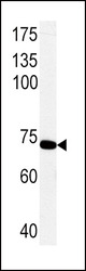
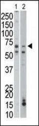
- Western blot analysis of PCK1 Antibody (C-term) (Cat. #AP8093b) in rat primary hepatocyte cell line lysates. PCK1 (arrow) was detected using the purified Pab.
- Primary Ab dilution
- 1:1000
- Submitted by
- Abcepta (provider)
- Main image
- Experimental details
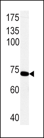
- PCK1 Antibody (C-term) (Cat. #AP8093b) western blot analysis in NCI-H460 cell line lysates (35ug/lane).This demonstrates the PCK1 antibody detected the PCK1 protein (arrow).
- Primary Ab dilution
- 1:1000

- Submitted by
- Abcepta (provider)
- Main image
- Experimental details
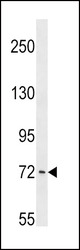
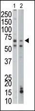
- The anti-PCK1 Pab (Cat. #AP8093b) is used in Western blot to detect PCK1 in mouse liver (Lane 1) and mouse small intestine tissue lysates (Lane 2).
- Primary Ab dilution
- 1:1000
Supportive validation
- Submitted by
- Abcepta (provider)
- Main image
- Experimental details
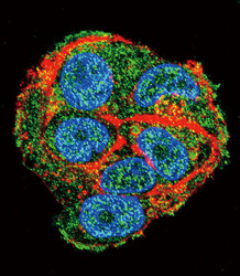
- Confocal immunofluorescent analysis of PCK1 Antibody (C-term)(Cat#AP8093b) with HepG2 cell followed by Alexa Fluor 488-conjugated goat anti-rabbit lgG (green). Actin filaments have been labeled with Alexa Fluor 555 phalloidin (red).DAPI was used to stain the cell nuclear (blue).
- Primary Ab dilution
- 1:10~50

Supportive validation
- Submitted by
- Abcepta (provider)
- Main image
- Experimental details
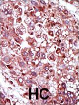
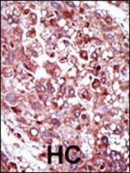
- "Formalin-fixed and paraffin-embedded human cancer tissue reacted with the primary antibody, which was peroxidase-conjugated to the secondary antibody, followed by AEC staining. This data demonstrates the use of this antibody for immunohistochemistry; clinical relevance has not been evaluated. BC = breast carcinoma; HC = hepatocarcinoma."
- Primary Ab dilution
- 1:50~100