 Explore
 Validate
 Learn
Learn Western blot
 Immunocytochemistry
Antibody data
- Antibody Data
- Antigen structure
- References [3]
- Comments [0]
- Validations
- Western blot [3]
- Immunohistochemistry [7]
Submit
Validation data
Reference
Comment
Report error
- Product number
- NBP1-86856 - Provider product page

- Provider
- Novus Biologicals
- Proper citation
- Novus Cat#NBP1-86856, RRID:AB_11019651
- Product name
- Rabbit Polyclonal WIRE Antibody
- Antibody type
- Polyclonal
- Description
- Immunogen affinity purified. Specificity of human, mouse, rat WIRE antibody verified on a Protein Array containing target protein plus 383 other non-specific proteins.
- Reactivity
- Human, Mouse, Rat
- Host
- Rabbit
- Isotype
- IgG
- Vial size
- 0.1 ml
- Storage
- Store at 4C short term. Aliquot and store at -20C long term. Avoid freeze-thaw cycles.
Submitted references A complex of ZO-1 and the BAR-domain protein TOCA-1 regulates actin assembly at the tight junction.
Cortical F-actin stabilization generates apical-lateral patterns of junctional contractility that integrate cells into epithelia.
N-WASP regulates the epithelial junctional actin cytoskeleton through a non-canonical post-nucleation pathway.
Van Itallie CM, Tietgens AJ, Krystofiak E, Kachar B, Anderson JM
Molecular biology of the cell 2015 Aug 1;26(15):2769-87
Molecular biology of the cell 2015 Aug 1;26(15):2769-87
Cortical F-actin stabilization generates apical-lateral patterns of junctional contractility that integrate cells into epithelia.
Wu SK, Gomez GA, Michael M, Verma S, Cox HL, Lefevre JG, Parton RG, Hamilton NA, Neufeld Z, Yap AS
Nature cell biology 2014 Feb;16(2):167-78
Nature cell biology 2014 Feb;16(2):167-78
N-WASP regulates the epithelial junctional actin cytoskeleton through a non-canonical post-nucleation pathway.
Kovacs EM, Verma S, Ali RG, Ratheesh A, Hamilton NA, Akhmanova A, Yap AS
Nature cell biology 2011 Jul 24;13(8):934-43
Nature cell biology 2011 Jul 24;13(8):934-43
No comments: Submit comment
Supportive validation
- Submitted by
- Novus Biologicals (provider)
- Main image
- Experimental details
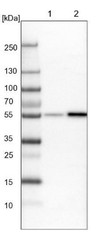
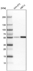
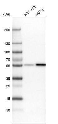
- Western Blot: WIRE Antibody [NBP1-86856] - Lane 1: NIH-3T3 cell lysate (Mouse embryonic fibroblast cells). Lane 2: NBT-II cell lysate (Rat Wistar bladder tumor cells).
- Submitted by
- Novus Biologicals (provider)
- Main image
- Experimental details
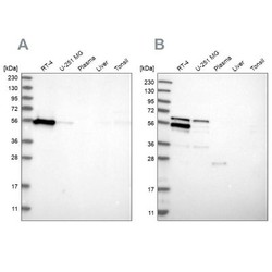
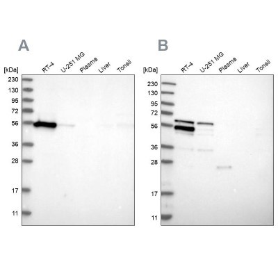
- Western Blot: WIRE Antibody [NBP1-86856] - Analysis using Anti-WIPF2 antibody NBP1-86856 (A) shows similar pattern to independent antibody NBP1-86858 (B).
- Submitted by
- Novus Biologicals (provider)
- Main image
- Experimental details
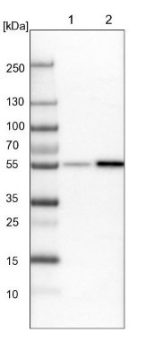
- Western Blot: WIRE Antibody [NBP1-86856] - Analysis in mouse cell line NIH-3T3 and rat cell line NBT-II.
Supportive validation
- Submitted by
- Novus Biologicals (provider)
- Main image
- Experimental details
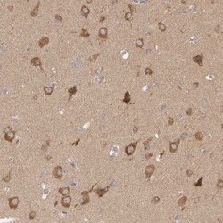
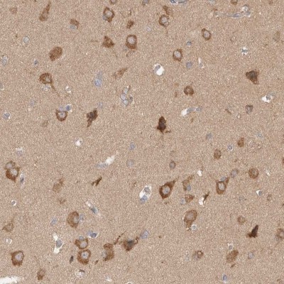
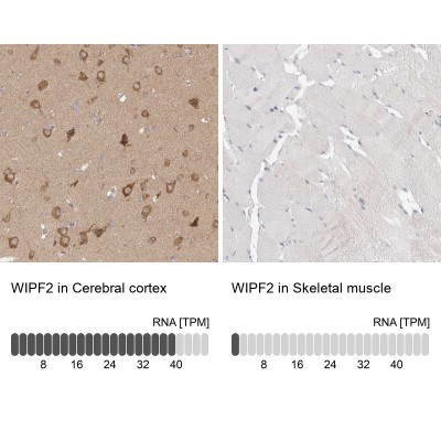
- Immunohistochemistry-Paraffin: WIRE Antibody [NBP1-86856] - Staining of human cerebral cortex shows high expression.
- Submitted by
- Novus Biologicals (provider)
- Main image
- Experimental details
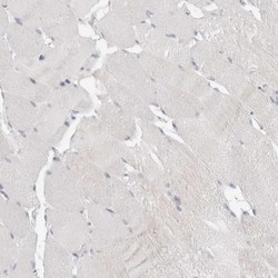
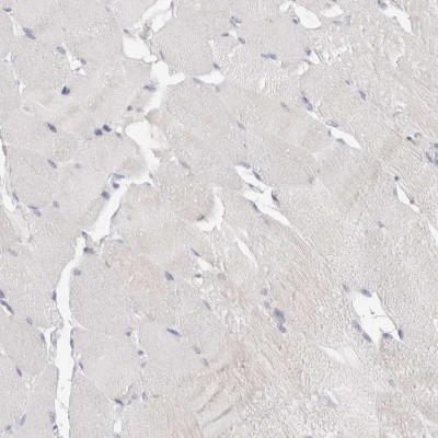
- Immunohistochemistry-Paraffin: WIRE Antibody [NBP1-86856] - Staining of human skeletal muscle shows low expression as expected.
- Submitted by
- Novus Biologicals (provider)
- Main image
- Experimental details
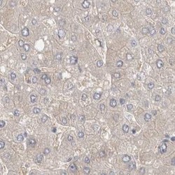
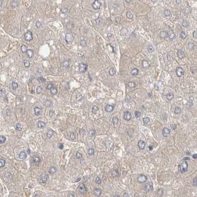
- Immunohistochemistry-Paraffin: WIRE Antibody [NBP1-86856] - Staining of human liver.
- Submitted by
- Novus Biologicals (provider)
- Main image
- Experimental details
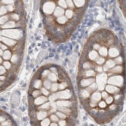
- Immunohistochemistry-Paraffin: WIRE Antibody [NBP1-86856] - Staining of human colon.

- Submitted by
- Novus Biologicals (provider)
- Main image

- Experimental details
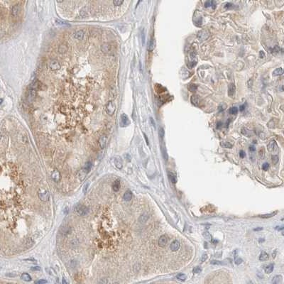
- Immunohistochemistry-Paraffin: WIRE Antibody [NBP1-86856] - Staining of human kidney.
- Submitted by
- Novus Biologicals (provider)
- Main image
- Experimental details
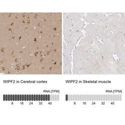
- Immunohistochemistry-Paraffin: WIRE Antibody [NBP1-86856] - Staining in human cerebral cortex and skeletal muscle tissues using anti-WIPF2 antibody. Corresponding WIPF2 RNA-seq data are presented for the same tissues.
- Submitted by
- Novus Biologicals (provider)
- Main image
- Experimental details
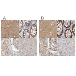
- Immunohistochemistry-Paraffin: WIRE Antibody [NBP1-86856] - Staining of human cerebral cortex, colon, kidney and liver using Anti-WIPF2 antibody NBP1-86856 (A) shows similar protein distribution across tissues to independent antibody NBP1-86858 (B).
