Explore
 Validate
 Learn
Learn Western blot
Antibody data
- Antibody Data
- Antigen structure
- References [0]
- Comments [0]
- Validations
- Western blot [6]
- Immunocytochemistry [5]
- Immunohistochemistry [1]
Submit
Validation data
Reference
Comment
Report error
- Product number
- PA5-77901 - Provider product page

- Provider
- Invitrogen Antibodies
- Product name
- Cdc6 Polyclonal Antibody
- Antibody type
- Polyclonal
- Antigen
- Recombinant full-length protein
- Description
- Positive Control: Jurkat, HeLa, HeLa (10 mM Hydroxyurea treatment for 18 hr), Neuro2A, GL261, C8D30, BCL-1, Raw264.7, C2C12, PC-12, Rat2
- Concentration
- 1.36 mg/mL
No comments: Submit comment
Supportive validation
- Submitted by
- Invitrogen Antibodies (provider)
- Main image
- Experimental details
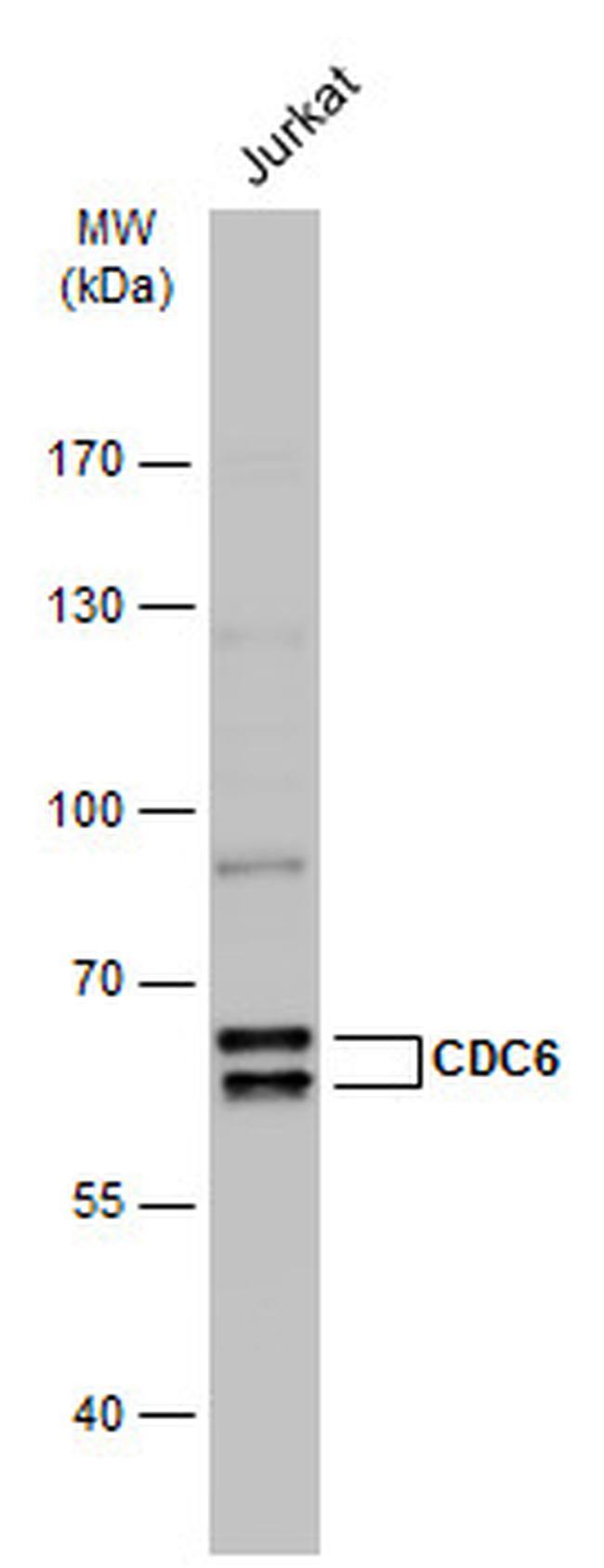
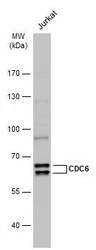
- Western blot analysis of Cdc6 in whole cell lysate using 30 µg of protein. Samples were separated with 7.5% SDS-PAGE and incubated with Cdc6 polyclonal antibody (Product # PA5-77901) using a dilution of 1:1000 followed by HRP-conjugated anti-rabbit IgG.
- Submitted by
- Invitrogen Antibodies (provider)
- Main image
- Experimental details
- Western blot analysis of Cdc6 in A) PC-12 whole cell lysate, B) Rat2 whole cell lysate using 30 µg of protein. Samples were separated with 7.5% SDS-PAGE and incubated with Cdc6 polyclonal antibody (Product # PA5-77901) using a dilution of 1:1000.

- Submitted by
- Invitrogen Antibodies (provider)
- Main image
- Experimental details
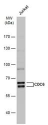
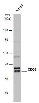
- Western Blot using Cdc6 Polyclonal Antibody (Product # PA5-77901). Whole cell extract (30 µg) was separated by 7.5% SDS-PAGE, and the membrane was blotted with Cdc6 Polyclonal Antibody (Product # PA5-77901) diluted at 1:1,000. The HRP-conjugated anti-rabbit IgG antibody was used to detect the primary antibody.
- Submitted by
- Invitrogen Antibodies (provider)
- Main image

- Experimental details
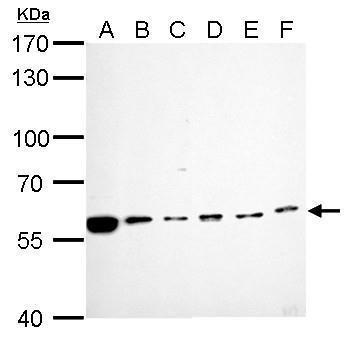
- Cdc6 Polyclonal Antibody detects CDC6 protein by western blot analysis. A. 30 µg Neuro2A whole cell lysate/extract. B. 30 µg GL261 whole cell lysate/extract. C. 30 µg C8D30 whole cell lysate/extract. D. 30 µg BCL-1 whole cell lysate/extract. E. 30 µg Raw264.7 whole cell lysate/extract. F. 30 µg C2C12 whole cell lysate/extract.7.5% SDS-PAGE. Cdc6 Polyclonal Antibody (Product # PA5-77901) dilution: 1:1,000. The HRP-conjugated anti-rabbit IgG antibody was used to detect the primary antibody.
- Submitted by
- Invitrogen Antibodies (provider)
- Main image

- Experimental details
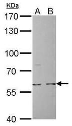
- Cdc6 Polyclonal Antibody detects CDC6 protein by western blot analysis. A. 30 µg PC-12 whole cell lysate/extract. B. 30 µg Rat2 whole cell lysate/extract.7.5% SDS-PAGE. Cdc6 Polyclonal Antibody (Product # PA5-77901) dilution: 1:1,000. The HRP-conjugated anti-rabbit IgG antibody was used to detect the primary antibody.

- Submitted by
- Invitrogen Antibodies (provider)
- Main image
- Experimental details
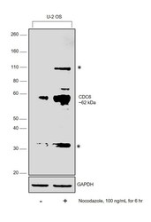
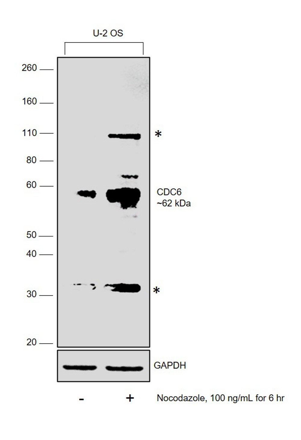
- Western Blot was performed using Anti-Cdc6 Polyclonal Antibody (Product # PA5-77901) and a 62 kDa band corresponding to Cell division control protein 6 homolog (CDC6) was upregulated upon nocodazole treatment in U-2 OS cells. Uncharacterised bands (*) at ~110 kDa and 32 kDa was also upregulated upon treatment. Whole cell extracts (40 µg lysate) of untreated U-2 OS (Lane 1) and nocodazole treated U-2 OS (Lane 2) were electrophoresed using NuPAGE™ 10% Bis-Tris Protein Gel (Product # NP0301BOX). Resolved proteins were then transferred onto a Nitrocellulose membrane (Product # IB23001) by iBlot® 2 Dry Blotting System (Product # IB21001). The Blot was probed with the primary antibody (1:2000 dilution) and detected by chemiluminescence with Goat anti-Rabbit IgG (H+L) Superclonal™ Recombinant Secondary Antibody, HRP (Product # A27036, 1:10000 dilution) using the iBright FL 1000 (Product # A32752). Chemiluminescent detection was performed using Novex® ECL Chemiluminescent Substrate Reagent Kit (Product # WP20005).
Supportive validation
- Submitted by
- Invitrogen Antibodies (provider)
- Main image

- Experimental details
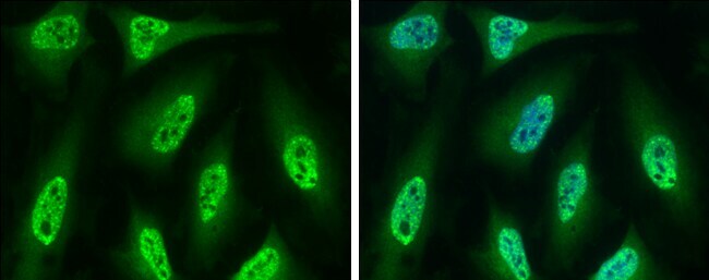
- Cdc6 Polyclonal Antibody detects CDC6 protein at nucleus by immunofluorescent analysis. Sample: HeLa cells were fixed in 4% paraformaldehyde at RT for 15 min. Green: CDC6 protein stained by Cdc6 Polyclonal Antibody (Product # PA5-77901) diluted at 1:500. Blue: Hoechst 33342 staining.
- Submitted by
- Invitrogen Antibodies (provider)
- Main image
- Experimental details
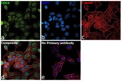
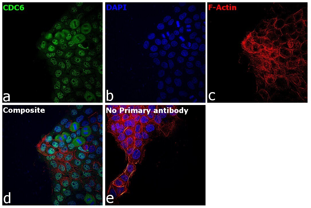
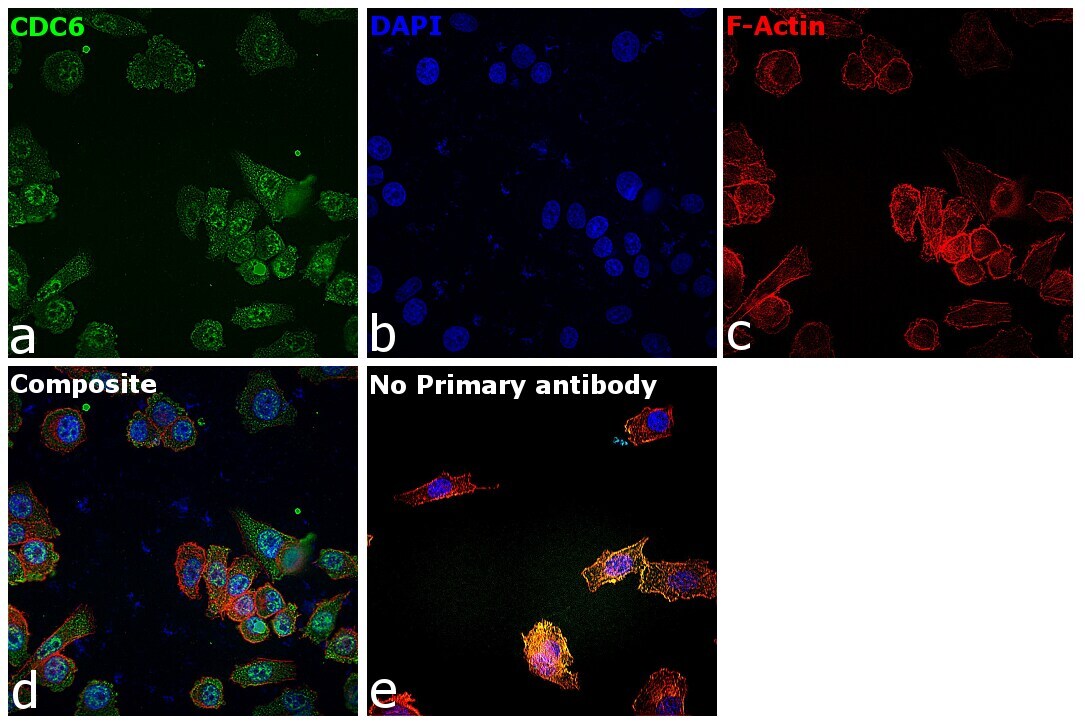
- Immunofluorescence analysis of CDC6 (Cell division control protein 6 homolog) was performed using 70% confluent log phase U-2 OS cells. The cells were fixed with 4% paraformaldehyde for 10 minutes, permeabilized with 0.1% Triton™ X-100 for 10 minutes, and blocked with 2% BSA for 45 minutes at room temperature. The cells were labeled with Cdc6 Polyclonal Antibody (Product # PA5-77901) at 1:100 in 0.1% BSA, incubated at 4 degree celsius overnight and then labeled with Donkey anti-Rabbit IgG (H+L) Highly Cross-Adsorbed Secondary Antibody, Alexa Fluor Plus 488 (Product # A32790), (1:2500), for 45 minutes at room temperature (Panel a: Green). Nuclei (Panel b:Blue) were stained with ProLong™ Diamond Antifade Mountant with DAPI (Product # P36962). F-actin (Panel c: Red) was stained with Rhodamine Phalloidin (Product # R415, 1:300). Panel d represents the merged image showing nuclear localization for CDC6. Panel e represents control cells with no primary antibody to assess background. The images were captured at 60X magnification.

- Submitted by
- Invitrogen Antibodies (provider)
- Main image

- Experimental details
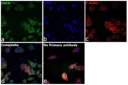
- Immunofluorescence analysis of CDC6 was performed using 70% confluent log phase A-431 cells. The cells were fixed with 4% paraformaldehyde for 10 minutes, permeabilized with 0.1% Triton™ X-100 for 15 minutes, and blocked with 2% BSA for 45 minutes at room temperature. The cells were labeled with Cdc6 Polyclonal Antibody (Product # PA5-77901) at 1:100 in 0.1% BSA, incubated at 4 degree celsius overnight and then labeled with Donkey anti-Rabbit IgG (H+L) Highly Cross-Adsorbed Secondary Antibody, Alexa Fluor Plus 488 (Product # A32790), (1:2000), for 45 minutes at room temperature (Panel a: Green). Nuclei (Panel b:Blue) were stained with Hoechst 33342 (Product # H1399). F-actin (Panel c: Red) was stained with Rhodamine Phalloidin (Product # R415, 1:300). Panel d represents the merged image showing predominant nuclear localization for most cells and cytoplasmic localisation in dividing cells for CDC6. Panel e represents control cells with no primary antibody to assess background. The images were captured at 40X magnification in CellInsight CX7 LZR High-Content Screening (HCS) Platform (Product # CX7C1115LZR).
- Submitted by
- Invitrogen Antibodies (provider)
- Main image
- Experimental details
- Immunofluorescence analysis of CDC6 was performed using 70% confluent log phase PC-3 cells. The cells were fixed with 4% paraformaldehyde for 10 minutes, permeabilized with 0.1% Triton™ X-100 for 15 minutes, and blocked with 2% BSA for 45 minutes at room temperature. The cells were labeled with Cdc6 Polyclonal Antibody (Product # PA5-77901) at 1:100 in 0.1% BSA, incubated at 4 degree celsius overnight and then labeled with Donkey anti-Rabbit IgG (H+L) Highly Cross-Adsorbed Secondary Antibody, Alexa Fluor Plus 488 (Product # A32790), (1:2000), for 45 minutes at room temperature (Panel a: Green). Nuclei (Panel b:Blue) were stained with Hoechst 33342 (Product # H1399). F-actin (Panel c: Red) was stained with Rhodamine Phalloidin (Product # R415, 1:300). Panel d represents the merged image showing predominant nuclear and cytoplasmic localisation for CDC6. Panel e represents control cells with no primary antibody to assess background. The images were captured at 40X magnification in CellInsight CX7 LZR High-Content Screening (HCS) Platform (Product # CX7C1115LZR).
- Submitted by
- Invitrogen Antibodies (provider)
- Main image

- Experimental details
- Immunofluorescence analysis of CDC6 was performed using 70% confluent log phase SH-SY5Y cells. The cells were fixed with 4% paraformaldehyde for 10 minutes, permeabilized with 0.1% Triton™ X-100 for 15 minutes, and blocked with 2% BSA for 45 minutes at room temperature. The cells were labeled with Cdc6 Polyclonal Antibody (Product # PA5-77901) at 1:100 in 0.1% BSA, incubated at 4 degree celsius overnight and then labeled with Donkey anti-Rabbit IgG (H+L) Highly Cross-Adsorbed Secondary Antibody, Alexa Fluor Plus 488 (Product # A32790), (1:2000), for 45 minutes at room temperature (Panel a: Green). Nuclei (Panel b:Blue) were stained with Hoechst 33342 (Product # H1399). F-actin (Panel c: Red) was stained with Rhodamine Phalloidin (Product # R415, 1:300). Panel d represents the merged image showing predominant nuclear localisation for CDC6. Panel e represents control cells with no primary antibody to assess background. The images were captured at 40X magnification in CellInsight CX7 LZR High-Content Screening (HCS) Platform (Product # CX7C1115LZR).
Supportive validation
- Submitted by
- Invitrogen Antibodies (provider)
- Main image
- Experimental details
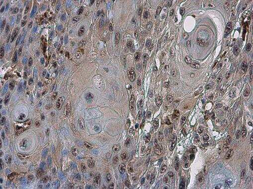
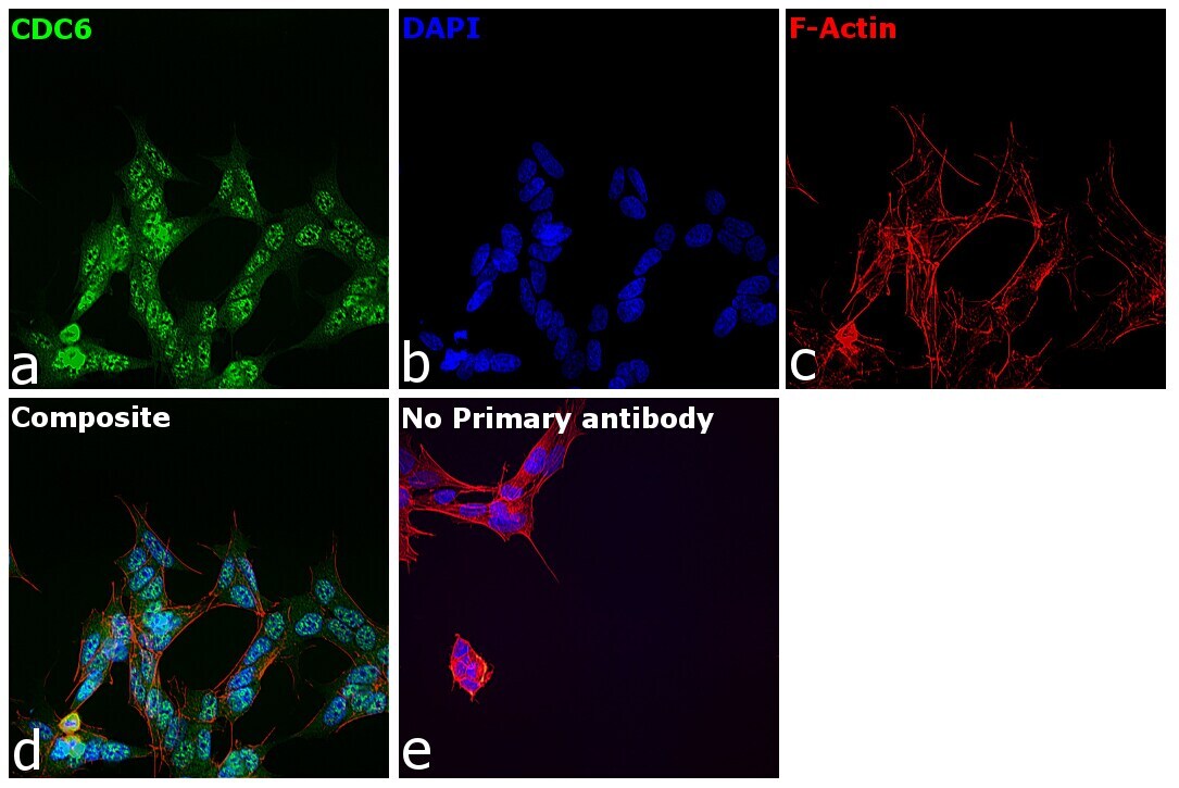
- Cdc6 Polyclonal Antibody detects CDC6 protein at cytoplasm in human oral carcinoma by immunohistochemical analysis. Sample: Paraffin-embedded human oral carcinoma. Cdc6 Polyclonal Antibody (Product # PA5-77901) diluted at 1:500. Antigen Retrieval: Citrate buffer, pH 6.0, 15 min.