 Explore
 Validate
 Learn
Learn Western blot
 Immunocytochemistry
Antibody data
- Antibody Data
- Antigen structure
- References [1]
- Comments [0]
- Validations
- Immunocytochemistry [1]
- Other assay [1]
Submit
Validation data
Reference
Comment
Report error
- Product number
- PA1-810A - Provider product page

- Provider
- Invitrogen Antibodies
- Product name
- RARA Polyclonal Antibody
- Antibody type
- Polyclonal
- Antigen
- Synthetic peptide
- Description
- PA1-810A detects retinoic acid receptor (RAR) from human and mouse tissues and cells. This antibody does not detect RAR beta or gamma forms. PA1-810A has been successfully used in Western blot procedures. By Western blot, this antibody detects an ~50-58 kDa protein representing RAR alpha from Jurkat, CEM-C7, and IM9 cells. The PA1-810A immunogen is a synthetic peptide corresponding to residues C S(444) P S L S P S S H R S S P A T Q S P (462) of mouse RAR alpha. PA1-810A immunizing peptide (Cat. # PEP-004) is available for use in neutralization and control experiments.
- Reactivity
- Human, Mouse
- Host
- Rabbit
- Isotype
- IgG
- Vial size
- 100 µL
- Concentration
- Conc. Not Determined
- Storage
- -20° C, Avoid Freeze/Thaw Cycles
Submitted references Neuroprotection against Amyloid-β-Induced DNA Double-Strand Breaks Is Mediated by Multiple Retinoic Acid-Dependent Pathways.
Colas J, Chessel N, Ouared A, Gruz-Gibelli E, Marin P, Herrmann FR, Savioz A
Neural plasticity 2020;2020:9369815
Neural plasticity 2020;2020:9369815
No comments: Submit comment
Supportive validation
- Submitted by
- Invitrogen Antibodies (provider)
- Main image
- Experimental details
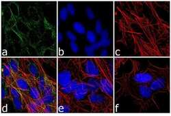
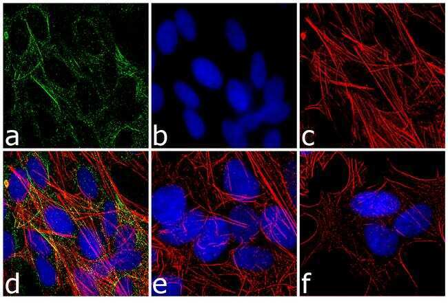
- Immunofluorescence analysis of Retinoic Acid Receptor alpha was performed using 70% confluent SH-SY5Y cells treated with 10µM retinoic acid for 72 hours. The cells were fixed with 4% paraformaldehyde for 10 minutes, permeabilized with 0.1% Triton™ X-100 for 10 minutes, and blocked with 1% BSA for 1 hour at room temperature. The cells were labeled with RARA Rabbit Polyclonal Antibody (Product # PA1-810A) at 1:250 dilution in 0.1% BSA and incubated for 3 hours at room temperature and then labeled with Goat anti-Rabbit IgG (H+L) Superclonal™ Secondary Antibody, Alexa Fluor® 488 conjugate (Product # A27034) at a dilution of 1:2000 for 45 minutes at room temperature (Panel a: green). Nuclei (Panel b: blue) were stained with SlowFade® Gold Antifade Mountant with DAPI (Product # S36938). F-actin (Panel c: red) was stained with Rhodamine Phalloidin (Product # R415, 1:300). Panel d represents the merged image showing cytoplasmic localization. Panel e is untreated cell with no signal. Panel f represents control cells with no primary antibody. The images were captured at 60X magnification.
Supportive validation
- Submitted by
- Invitrogen Antibodies (provider)
- Main image
- Experimental details
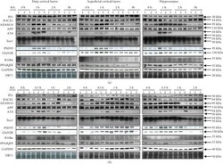
- Figure 3 Changes in protein expression following 1 mu M all- trans -retinoic acid (RA) treatment of the deep and superficial neocortical layers, as well as of the hippocampus, of (a) three 1-month-old and (b) three 17-month-old male C57BL/6J mice. The proteins are involved in the amyloid cascade (Presenilin 1 or PS1/ gamma -secretase, BACE1/ beta -secretase, ADAM10/ alpha -secretase, and APP C-terminus), in Tau phosphorylation (phospho-Tau (AT8) or unphosphorylated Tau (Tau1)), in synaptic functions (PSD95, GluN2B/NR2B), or in RA-dependent pathways (RAR alpha , PPAR beta / delta ). GAPDH and DB71 stainings were used to demonstrate equal loading of the Western blot gels. Overall, we observed significant increases (see Section 3.3 ) of ADAM10, APP, and phosphorylated and unphosphorylated Tau proteins, suggesting the activation of neuroprotective mechanisms following the RA treatment, whereas the expression of the enzymes of the amyloidogenic pathway, PS1 and BACE1, or, in most cases, of the RA receptors was not increased. Protein sizes are indicated on the right. A size range is given for Tau isoforms (AT8 and Tau1).
