 Explore
 Validate
 Learn
Learn Western blot
Antibody data
- Antibody Data
- Antigen structure
- References [1]
- Comments [0]
- Validations
- Western blot [1]
- Immunocytochemistry [2]
- Flow cytometry [1]
Submit
Validation data
Reference
Comment
Report error
- Product number
- MAB83771-100 - Provider product page

- Provider
- R&D Systems
- Product name
- Human/Mouse/Rat HHEX Antibody
- Antibody type
- Monoclonal
- Description
- Protein A or G purified from hybridoma culture supernatant. Detects human HHEX in direct ELISAs and Western blots.
- Reactivity
- Human, Mouse, Rat
- Host
- Rabbit
- Conjugate
- Unconjugated
- Antigen sequence
Q03014- Isotype
- IgG
- Antibody clone number
- 2018B
- Vial size
- 100 ug
- Storage
- Use a manual defrost freezer and avoid repeated freeze-thaw cycles. 12 months from date of receipt, -20 to -70 °C as supplied. 1 month, 2 to 8 °C under sterile conditions after reconstitution. 6 months, -20 to -70 °C under sterile conditions after reconstitution.
Submitted references Single-Cell RNA-Sequencing-Based CRISPRi Screening Resolves Molecular Drivers of Early Human Endoderm Development.
Genga RMJ, Kernfeld EM, Parsi KM, Parsons TJ, Ziller MJ, Maehr R
Cell reports 2019 Apr 16;27(3):708-718.e10
Cell reports 2019 Apr 16;27(3):708-718.e10
No comments: Submit comment
Supportive validation
- Submitted by
- R&D Systems (provider)
- Main image
- Experimental details
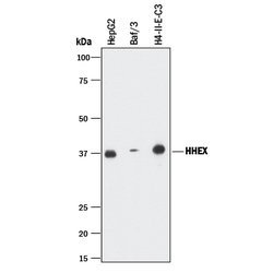
- Detection of Human, Mouse, and Rat HHEX by Western Blot. Western blot shows lysates of HepG2 human hepatocellular carcinoma cell line, BaF3 mouse pro-B cell line, and H4-II-E-C3 rat hepatoma cell line. PVDF membrane was probed with 1 µg/mL of Rabbit Anti-Human/Mouse/Rat HHEX Monoclonal Antibody (Catalog # MAB83771) followed by HRP-conjugated Anti-Rabbit IgG Secondary Antibody (Catalog # HAF008). A specific band was detected for HHEX at approximately 37 kDa (as indicated). This experiment was conducted under reducing conditions and using Immunoblot Buffer Group 1.

Supportive validation
- Submitted by
- R&D Systems (provider)
- Main image
- Experimental details
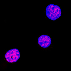
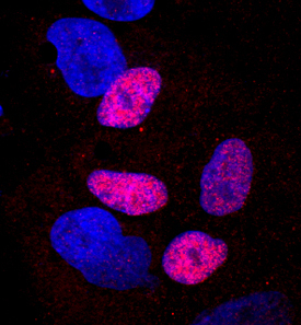
- HHEX in K562 Human Cell Line. HHEX was detected in immersion fixed K562 human chronic myelogenous leukemia cell line using Rabbit Anti-Human/Mouse/Rat HHEX Polyclonal Antibody (Catalog # MAB83771) at 3 µg/mL for 3 hours at room temperature. Cells were stained using the NorthernLights™ 557-conjugated Anti-Rabbit IgG Secondary Antibody (red; Catalog # NL004) and counterstained with DAPI (blue). Specific staining was localized to nuclei. View our protocol for Fluorescent ICC Staining of Non-adherent Cells.
- Submitted by
- R&D Systems (provider)
- Main image

- Experimental details
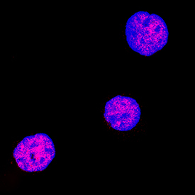
- HHEX in iBJ6 iPS Cell Line. HHEX was detected in immersion fixed iBJ6 iPS cell line differentiated into hepatocytes using Rabbit Anti-Human/Mouse/Rat HHEX Polyclonal Antibody (Catalog # MAB83771) at 2 µg/mL for 3 hours at room temperature. Cells were stained using the NorthernLights™ 557-conjugated Anti-Rabbit IgG Secondary Antibody (red; Catalog # NL004) and counterstained with DAPI (blue). Specific staining was localized to nuclei. View our protocol for Fluorescent ICC Staining of Cells on Coverslips.
Supportive validation
- Submitted by
- R&D Systems (provider)
- Main image
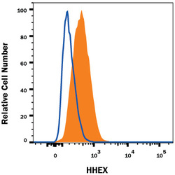
- Experimental details
- Detection of HHEX in A549 Human Cell Line by Flow Cytometry. A549 human lung carcinoma cell line was stained with Rabbit Anti-Human/Mouse/Rat HHEX Monoclonal Antibody (Catalog # MAB83771, filled histogram) or isotype control antibody (Catalog # AB-105-C, open histogram), followed by Phycoerythrin-conjugated Anti-Rabbit IgG Secondary Antibody (Catalog # F0110). To facilitate intracellular staining, cells were fixed and permeabilized with FlowX FoxP3 Fixation & Permeabilization Buffer Kit (Catalog # FC012). View our protocol for Staining Intracellular Molecules.
