Explore
 Validate
 Learn
Learn Western blot
Antibody data
- Antibody Data
- Antigen structure
- References [1]
- Comments [0]
- Validations
- Western blot [2]
- Other assay [1]
Submit
Validation data
Reference
Comment
Report error
- Product number
- PA5-19625 - Provider product page

- Provider
- Invitrogen Antibodies
- Product name
- Hex Polyclonal Antibody
- Antibody type
- Polyclonal
- Antigen
- Synthetic peptide
- Description
- For Western Blot, this antibody has non-specific bands at 42 kDa.
- Concentration
- 0.8 mg/mL
Submitted references Developmental kinetics and transcriptome dynamics of stem cell specification in the spermatogenic lineage.
Law NC, Oatley MJ, Oatley JM
Nature communications 2019 Jun 26;10(1):2787
Nature communications 2019 Jun 26;10(1):2787
No comments: Submit comment
Supportive validation
- Submitted by
- Invitrogen Antibodies (provider)
- Main image
- Experimental details
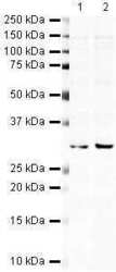
- Western blot analysis of HeLa Whole Cell Lysate using Product # PA5-19625, Hex primary antibody at a dilution of 1:250 (lane 1). Staining of HEK 293 Whole Cell Lysate at a dilution of 1:250 (lane 2). Blot treated with a secondary IR Dye680-conjugated Goat polyclonal anti-Rabbit antibody was used at a dilution of 1:10000.

- Submitted by
- Invitrogen Antibodies (provider)
- Main image

- Experimental details
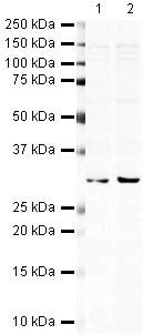
- Western blot analysis of HeLa Whole Cell Lysate using Product # PA5-19625, Hex primary antibody at a dilution of 1:250 (lane 1). Staining of HEK 293 Whole Cell Lysate at a dilution of 1:250 (lane 2). Blot treated with a secondary IR Dye680-conjugated Goat polyclonal anti-Rabbit antibody was used at a dilution of 1:10000.
Supportive validation
- Submitted by
- Invitrogen Antibodies (provider)
- Main image
- Experimental details
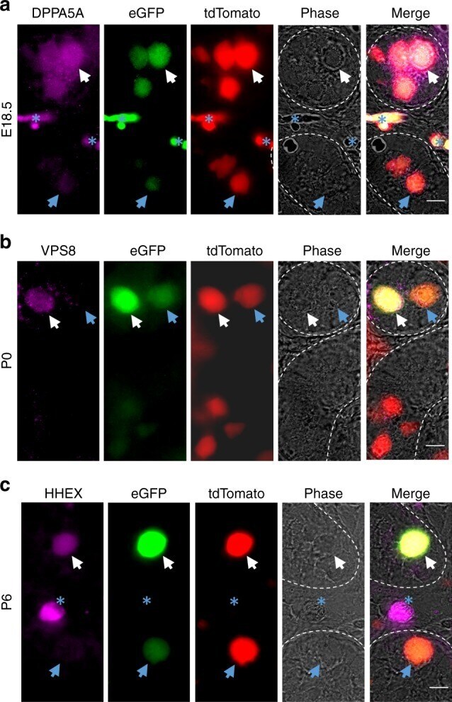
- Fig. 5 Validation of markers of SSC fate specification identified by scRNA-seq clustering. a - c Immunofluorescent staining for DPPA5A ( a ), VPS8 ( b ), and HHEX ( c ) proteins that were identified as differentially expressed at the transcript level in germ cell clusters 1, 5, and 7 of E16.5, P0, and P6 testes by scRNA-seq, respectively. Immunostaining is overlaid with ID4-eGFP and tdTomato fluorescence in germ cells. White arrows indicate germ cells that are DPPA5A+ and ID4-eGFP+ at E18.5, VPS8+ and ID4-eGFP Bright at P0, or HHEX+ and ID4-eGFP Bright at P6. Blue arrows indicate germ cells that have low to undetectable staining for the selected marker and ID4-eGFP. Blue asterisks denote in ( a ) vasculature in E18.5 testes that has autofluorescence, and in ( c ) an interstitial cell (i.e. tdTomato-) that is HHEX+. Seminiferous tubule borders are indicated by with white dotted lines. Scale bars, 10 um. Images are representative of >=3 cross-sections imaged from n = 2 biologically independent animals for each developmental age point