Explore
 Validate
 Learn
Learn Western blot
Antibody data
- Antibody Data
- Antigen structure
- References [0]
- Comments [0]
- Validations
- Western blot [3]
- Immunocytochemistry [4]
Submit
Validation data
Reference
Comment
Report error
- Product number
- NBP2-29982 - Provider product page

- Provider
- Novus Biologicals
- Product name
- Mouse Monoclonal Fatty Acid Synthase/FASN Antibody
- Antibody type
- Monoclonal
- Description
- Ammonium sulfate precipitation.
- Reactivity
- Human, Mouse
- Host
- Mouse
- Isotype
- IgG
- Vial size
- 0.4 ml
- Concentration
- 7.8 mg/ml
- Storage
- Store at 4C short term. Aliquot and store at -20C long term. Avoid freeze-thaw cycles.
No comments: Submit comment
Supportive validation
- Submitted by
- Novus Biologicals (provider)
- Main image

- Experimental details
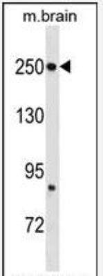
- Western Blot: Fatty Acid Synthase/FASN Antibody (497CT15.2.5) [NBP2-29982] - Western blot analysis in mouse brain tissue lysates (35ug/lane).This demonstrates the FASN (Center) antibody detected the FASN (Center) protein (arrow).
- Submitted by
- Novus Biologicals (provider)
- Main image

- Experimental details
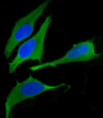
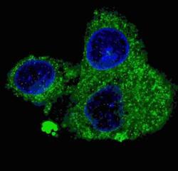
- Western Blot: Fatty Acid Synthase/FASN Antibody (497CT15.2.5) [NBP2-29982] - Fluorescent confocal image of HepG2 cells stained with FASN (Center) antibody. HepG2 cells were fixed with 4% PFA (20 min), permeabilized with Triton X-100 (0.2%, 30 min). Cells were then incubated with AM2067b FASN primary antibody (1:200, 2 h at room temperature). For secondary antibody, Alexa Fluor 488 conjugated donkey anti-mouse antibody (green) was used (1:1000, 1h). Nuclei were counterstained with Hoechst 33342 (blue) (10ug/ml, 5 min). Note the highly specific localization of the FASN immunosignal to the cytoplasm, supported by Human Protein Atlas Data.
- Submitted by
- Novus Biologicals (provider)
- Main image
- Experimental details
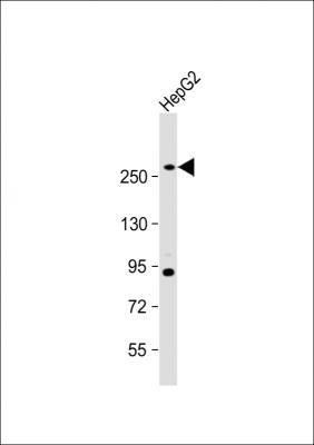
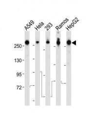
- Western Blot: Fatty Acid Synthase/FASN Antibody (497CT15.2.5) [NBP2-29982] - All lanes : Anti-FASN Antibody (Center). Lane 1: A549 whole cell lysate; Lane 2: Hela whole cell lysate; Lane 3: 293 whole cell lysate; Lane 4: Ramos whole cell lysate; Lane 5: HepG2 whole cell lysate. Lysates/proteins at 20 ug per lane. Secondary Goat Anti-mouse IgG, (H+L), Peroxidase conjugated. Predicted band size : 273 kDa. Blocking/Dilution buffer: 5% NFDM/TBST.

Supportive validation
- Submitted by
- Novus Biologicals (provider)
- Main image
- Experimental details
- Immunocytochemistry/Immunofluorescence: Fatty Acid Synthase/FASN Antibody (497CT15.2.5) [NBP2-29982] - Fluorescent confocal image of HepG2 cells stained with FASN (Center) antibody. HepG2 cells were fixed with 4% PFA (20 min), permeabilized with Triton X-100 (0.2%, 30 min). Cells were then incubated with AM2067b FASN primary antibody (1:200, 2 h at room temperature). For secondary antibody, Alexa Fluor 488 conjugated donkey anti-mouse antibody (green) was used (1:1000, 1h). Nuclei were counterstained with Hoechst 33342 (blue) (10 ug/ml, 5 min). Note the highly specific localization of the FASN immunosignal to the cytoplasm, supported by Human Protein Atlas Data

- Submitted by
- Novus Biologicals (provider)
- Main image
- Experimental details
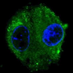
- Immunocytochemistry/Immunofluorescence: Fatty Acid Synthase/FASN Antibody (497CT15.2.5) [NBP2-29982] - Analysis using Unpurified version of NBP2-29982. Fluorescent confocal image of HepG2 cells stained with FASN (Center) antibody. HepG2 cells were fixed with 4% PFA (20 min), permeabilized with Triton X-100 (0.2%, 30 min).
- Submitted by
- Novus Biologicals (provider)
- Main image
- Experimental details
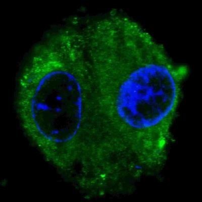
- Immunofluorescence: Fatty Acid Synthase/FASN Antibody (497CT15.2.5) [NBP2-29982] - Immunofluorescent analysis of 4% paraformaldehyde-fixed, 0. 1% Triton X-100 permeabilized HepG2 (human liver hepatocellular carcinoma cell line) cells labeling FASN with NBP2-29982, followed by Dylight(R) 488-conjugated goat anti-mouse IgG secondary antibody (green). Immunofluorescence image showing cytoplasm HepG2 cell line. The nuclear counter stain is DAPI (blue).

- Submitted by
- Novus Biologicals (provider)
- Main image

- Experimental details
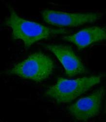
- Immunofluorescence: Fatty Acid Synthase/FASN Antibody (497CT15.2.5) [NBP2-29982] - Immunofluorescent analysis of 4% paraformaldehyde-fixed, 0. 1% Triton X-100 permeabilized U-2 OS ((human cervical epithelial adenocarcinoma cell line) cells labeling FASN with NBP2-29982, followed by Dylight(R) 488-conjugated goat anti-mouse IgG secondary antibody (green). Immunofluorescence image showing cytoplasm Hela cell line. The nuclear counter stain is DAPI (blue).