 Explore
 Validate
 Learn
Learn Western blot
 Immunocytochemistry
Antibody data
- Antibody Data
- Antigen structure
- References [1]
- Comments [0]
- Validations
- Immunocytochemistry [2]
- Immunoprecipitation [1]
- Immunohistochemistry [5]
- Other assay [2]
Submit
Validation data
Reference
Comment
Report error
- Product number
- PA5-22061 - Provider product page

- Provider
- Invitrogen Antibodies
- Product name
- FASN Polyclonal Antibody
- Antibody type
- Polyclonal
- Antigen
- Recombinant full-length protein
- Description
- Recommended positive controls: Molt-4, mouse brain, PC-12, Rat2, 293T. Predicted reactivity: Mouse (89%), Rat (89%), Pig (83%), Bovine (85%). Store product as a concentrated solution. Centrifuge briefly prior to opening the vial.
- Reactivity
- Human, Mouse, Rat
- Host
- Rabbit
- Isotype
- IgG
- Vial size
- 100 μL
- Concentration
- 0.2 mg/mL
- Storage
- Store at 4°C short term. For long term storage, store at -20°C, avoiding freeze/thaw cycles.
Submitted references Antiobesity and antidiabetic effects of the dairy bacterium Propionibacterium freudenreichii MJ2 in high-fat diet-induced obese mice by modulating lipid metabolism.
An M, Park YH, Lim YH
Scientific reports 2021 Jan 28;11(1):2481
Scientific reports 2021 Jan 28;11(1):2481
No comments: Submit comment
Supportive validation
- Submitted by
- Invitrogen Antibodies (provider)
- Main image
- Experimental details
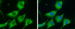
- Immunocytochemistry-Immunofluorescence analysis of FASN was performed in HeLa cells fixed in 4% paraformaldehyde at RT for 15 min. Green: FASN Polyclonal Antibody (Product # PA5-22061) diluted at 1:500. Blue: Hoechst 33342 staining.

- Submitted by
- Invitrogen Antibodies (provider)
- Main image

- Experimental details
- Immunocytochemistry-Immunofluorescence analysis of FASN was performed in HeLa cells fixed in 4% paraformaldehyde at RT for 15 min. Green: FASN Polyclonal Antibody (Product # PA5-22061) diluted at 1:500. Blue: Hoechst 33342 staining.

Supportive validation
- Submitted by
- Invitrogen Antibodies (provider)
- Main image

- Experimental details
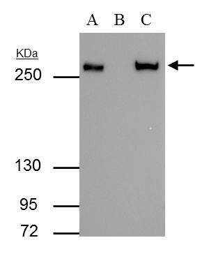
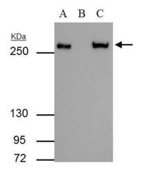
- FASN antibody immunoprecipitates FASN protein in IP experiments. IP Sample: HeLa whole cell lysate/extract A : 30 µg whole cell lysate/extract of FASN protein expressing HeLa cells B : Control with 2.5 µg of pre-immune rabbit IgG C : Immunoprecipitation of FASN by 2.5 µg of FASN antibody (Product # PA5-22061) 5% SDS-PAGE The immunoprecipitated FASN protein was detected by FASN antibody (Product # PA5-22061) diluted at 1:1,000. Anti-rabbit IgG (HRP) was used as a secondary reagent.
Supportive validation
- Submitted by
- Invitrogen Antibodies (provider)
- Main image
- Experimental details
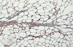
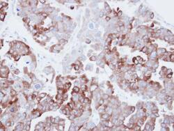
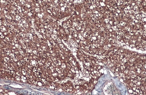
- FASN Polyclonal Antibody detects Fatty Acid Synthase protein at cytoplasm by immunohistochemical analysis. Sample: Paraffin-embedded mouse white adipocyte. Fatty Acid Synthase stained by FASN Polyclonal Antibody (Product # PA5-22061) diluted at 1:500. Antigen Retrieval: Citrate buffer, pH 6.0, 15 min.
- Submitted by
- Invitrogen Antibodies (provider)
- Main image

- Experimental details
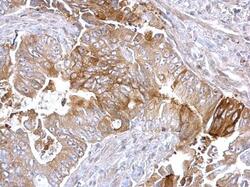
- FASN Polyclonal Antibody detects Fatty Acid Synthase protein at cytoplasm on human breast carcinoma by immunohistochemical analysis. Sample: Paraffin-embedded human breast carcinoma. FASN Polyclonal Antibody (Product # PA5-22061) diluted at 1:500. Antigen Retrieval: EDTA based buffer, pH 8.0, 15 min.
- Submitted by
- Invitrogen Antibodies (provider)
- Main image
- Experimental details
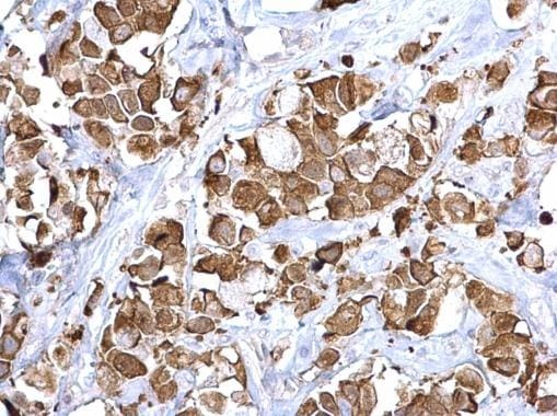
- FASN Polyclonal Antibody detects Fatty Acid Synthase protein at cytoplasm on human colon carcinoma by immunohistochemical analysis. Sample: Paraffin-embedded human colon carcinoma. FASN Polyclonal Antibody (Product # PA5-22061) diluted at 1:500. Antigen Retrieval: EDTA based buffer, pH 8.0, 15 min.

- Submitted by
- Invitrogen Antibodies (provider)
- Main image
- Experimental details
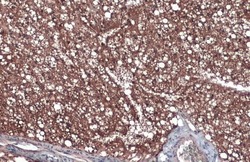
- Immunohistochemical analysis of paraffin-embedded human ovarian carainoma, using Fatty Acid Synthase (Product # PA5-22061) antibody at 1:500 dilution. Antigen Retrieval: EDTA based buffer, pH 8.0, 15 min.

- Submitted by
- Invitrogen Antibodies (provider)
- Main image
- Experimental details
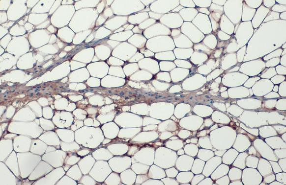
- FASN Polyclonal Antibody detects Fatty Acid Synthase protein at cytoplasm by immunohistochemical analysis. Sample: Paraffin-embedded mouse brown adipocyte. Fatty Acid Synthase stained by FASN Polyclonal Antibody (Product # PA5-22061) diluted at 1:500. Antigen Retrieval: Citrate buffer, pH 6.0, 15 min.
Supportive validation
- Submitted by
- Invitrogen Antibodies (provider)
- Main image
- Experimental details
- FASN antibody immunoprecipitates FASN protein in IP experiments. IP Sample: HeLa whole cell lysate/extract A : 30 µg whole cell lysate/extract of FASN protein expressing HeLa cells B : Control with 2.5 µg of pre-immune rabbit IgG C : Immunoprecipitation of FASN by 2.5 µg of FASN antibody (Product # PA5-22061) 5% SDS-PAGE The immunoprecipitated FASN protein was detected by FASN antibody (Product # PA5-22061) diluted at 1:1,000. Anti-rabbit IgG (HRP) was used as a secondary reagent.

- Submitted by
- Invitrogen Antibodies (provider)
- Main image

- Experimental details
- Figure 5 Effects of MJ2 and hkMJ2 on protein expression levels related to adipogenesis and lipid metabolism in eWAT. Total protein was extracted from eWAT in each group, and the relative protein expression levels of the factors related to ( A ) adipogenesis (PPARgamma and C/EBPalpha), ( B ) lipogenesis (FAS, SCD-1 and ACC) and ( C ) lipolysis (ATGL and HSL) were investigated by western blot. Representative images of each protein are shown, and the relative quantified expression levels indicate the mean +- SD. The p values were determined by ANOVA and Tukey's HSD test. The full-length blots are shown in Supplementary Fig. 3 .
