Explore
 Validate
 Learn
Learn Western blot
 Immunocytochemistry
Antibody data
- Antibody Data
- Antigen structure
- References [1]
- Comments [0]
- Validations
- Western blot [1]
- Immunocytochemistry [1]
- Immunohistochemistry [1]
Submit
Validation data
Reference
Comment
Report error
- Product number
- HPA012024 - Provider product page

- Provider
- Atlas Antibodies
- Proper citation
- Atlas Antibodies Cat#HPA012024, RRID:AB_1846503
- Product name
- Anti-CEBPG
- Antibody type
- Polyclonal
- Description
- Polyclonal Antibody against Human CEBPG, Gene description: CCAAT/enhancer binding protein (C/EBP), gamma, Alternative Gene Names: GPE1BP, IG/EBP-1, Validated applications: ICC, IHC, WB, Uniprot ID: P53567, Storage: Store at +4°C for short term storage. Long time storage is recommended at -20°C.
- Reactivity
- Human
- Host
- Rabbit
- Conjugate
- Unconjugated
- Isotype
- IgG
- Vial size
- 100 µl
- Concentration
- 0.1 mg/ml
- Storage
- Store at +4°C for short term storage. Long time storage is recommended at -20°C.
- Handling
- The antibody solution should be gently mixed before use.
Submitted references Activating transcription factor 3 coordinates differentiation of cardiac and hematopoietic progenitors by regulating glucose metabolism
Yin H, Yan L, Liu Q, Peng Z, Zhang C, Xia Y, Su D, Gu A, Zhou Y
Science Advances 2020;6(19)
Science Advances 2020;6(19)
No comments: Submit comment
Enhanced validation
- Submitted by
- Atlas Antibodies (provider)
- Enhanced method
- Recombinant expression validation
- Main image

- Experimental details
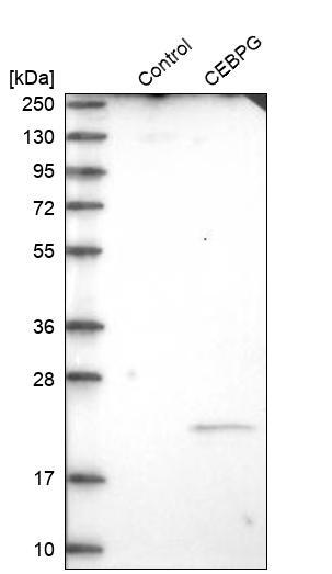
- Western blot analysis in control (vector only transfected HEK293T lysate) and CEBPG over-expression lysate (Co-expressed with a C-terminal myc-DDK tag (~3.1 kDa) in mammalian HEK293T cells, LY400686).
- Sample type
- Human
- Protocol
- Protocol
Supportive validation
- Submitted by
- Atlas Antibodies (provider)
- Main image
- Experimental details
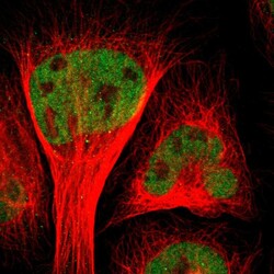
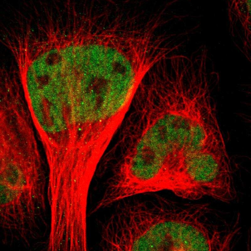
- Immunofluorescent staining of human cell line U-2 OS shows localization to nucleoplasm.
- Sample type
- Human
Supportive validation
- Submitted by
- Atlas Antibodies (provider)
- Enhanced method
- Orthogonal validation
- Main image

- Experimental details
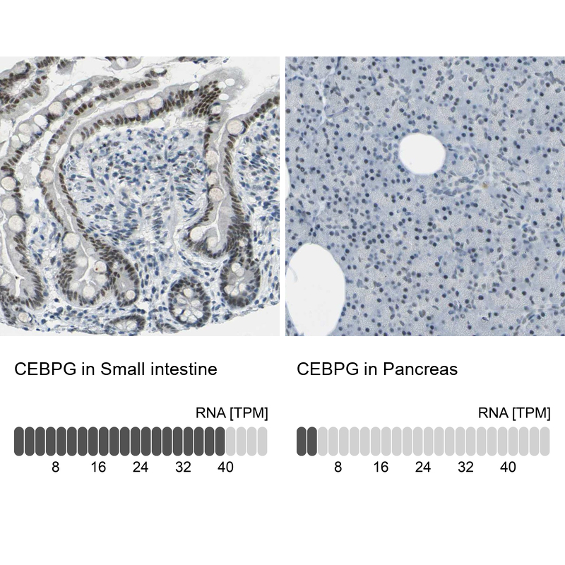
- Immunohistochemistry analysis in human small intestine and pancreas tissues using Anti-CEBPG antibody. Corresponding CEBPG RNA-seq data are presented for the same tissues.
- Sample type
- Human
- Protocol
- Protocol