 Explore
 Validate
 Learn
Learn Immunohistochemistry
Antibody data
- Antibody Data
- Antigen structure
- References [0]
- Comments [0]
- Validations
- Immunohistochemistry [2]
- Other assay [1]
Submit
Validation data
Reference
Comment
Report error
- Product number
- 50-9015-82 - Provider product page

- Provider
- Invitrogen Antibodies
- Product name
- Phospho-BTK/ITK (Tyr551, Tyr511) Monoclonal Antibody (M4G3LN), eFluor™ 660, eBioscience™
- Antibody type
- Monoclonal
- Antigen
- Other
- Description
- Description: This M4G3LN monoclonal antibody recognizes human and mouse Bruton's tyrosine kinase (also known as BTK, agammaglobulinaemia tyrosine kinase, B-cell progenitor kinase)/interleukin-2-inducible tyrosine kinase (also known as ITK, T-cell-specific kinase, Tyrosine-protein kinase Lyk) when phosphorylated on Y551 and Y511, respectively. In addition to Src family and SYK family kinases, B cells and T cells express a third family of receptor-proximal kinases called the Tec kinase family. The B cell-specific Tec family kinase is BTK; T cells express ITK as well as Tec and Rlk. Tec family kinases play an indispensable role in activation of phospholipase C and activation of calcium-dependent signaling cascades following antigen receptor triggering. BTK has been implicated in propagating signals downstream of LPS-TLR4 in B cells via signaling through the TLR4 co-receptor RP105/CD180. Mice congenitally lacking BTK or ITK show profound defects in B cell or T cell development and signaling, respectively. People with mutations or deletions in BTK suffer from X-linked agammaglobulinemia and X-linked immunodeficiency. Specificity of this M4G3LN clone was determined by ELISA, flow cytometry, and western blotting. Applications Reported: This M4G3LN antibody has been reported for use in immunohistochemical staining, and immunohistochemical staining of formalin-fixed paraffin embedded tissue sections. Applications Tested: This M4G3LN antibody has been tested by immunohistochemistry of formalin-fixed paraffin embedded human tissue using high pH antigen retrieval and can be used at less than or equal to10 µg/mL. It is recommended that the antibody be carefully titrated for optimal performance in the assay of interest. eFluor® 660 is a replacement for Alexa Fluor® 647. eFluor® 660 emits at 659 nm and is excited with the red laser (633 nm). Please make sure that your instrument is capable of detecting this fluorochome. Excitation: 633-647 nm; Emission: 668 nm; Laser: Red Laser. Filtration: 0.2 µm post-manufacturing filtered.
- Reactivity
- Human, Mouse
- Host
- Mouse
- Isotype
- IgG
- Antibody clone number
- M4G3LN
- Vial size
- 100 µg
- Concentration
- 0.2 mg/mL
- Storage
- 4° C, store in dark, DO NOT FREEZE!
No comments: Submit comment
Supportive validation
- Submitted by
- Invitrogen Antibodies (provider)
- Main image
- Experimental details
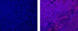
- Immunohistochemistry of formalin-fixed paraffin embedded human tonsil tissue using 10 µg/mL Mouse IgG2b K Isotype Control eFluor® 660 (left) or 10 µg/mL Anti-Human/Mouse phospho-BTK/ITK (Y551/Y511) eFluor® 660 (right). Nuclei are stained with DAPI.

- Submitted by
- Invitrogen Antibodies (provider)
- Main image

- Experimental details
- Immunohistochemistry of formalin-fixed paraffin embedded human tonsil tissue using 10 µg/mL Mouse IgG2b K Isotype Control eFluor® 660 (left) or 10 µg/mL Anti-Human/Mouse phospho-BTK/ITK (Y551/Y511) eFluor® 660 (right). Nuclei are stained with DAPI.

Supportive validation
- Submitted by
- Invitrogen Antibodies (provider)
- Main image

- Experimental details
- Figure 8 Enrichment and activation of B cell-associated signaling pathways in HS skin. Analysis of the signal transduction networks using literature-based networks (Genomatix-Pathway System, GePS) demonstrated enrichment for pathways involved in B cell signaling and activation ( A ). To confirm the nature of the inflammatory infiltrate in HS and the localization of components of the enriched signaling pathways, we performed IHC in an excisional biopsy for CD3, CD20, and CD138. Plasma cells were the predominant inflammatory infiltrate and most prominent in the deeper layers of the skin surrounding a deeper sinus tract ( A ), accompanied by increased expression of BTK, SYK, and LCK ( B ) ( n = 3). Activation of key components of this signaling pathway was confirmed by IHC for both phospho-BTK and phospho-SYK ( n = 3) ( C ).
