Explore
 Validate
 Learn
Learn Western blot
Antibody data
- Antibody Data
- Antigen structure
- References [1]
- Comments [0]
- Validations
- Western blot [1]
- Immunocytochemistry [2]
- Immunohistochemistry [1]
Submit
Validation data
Reference
Comment
Report error
- Product number
- GTX103592 - Provider product page

- Provider
- GeneTex
- Proper citation
- GeneTex Cat#GTX103592, RRID:AB_1952476
- Product name
- UAP1 antibody [N1C3]
- Antibody type
- Polyclonal
- Reactivity
- Human
- Host
- Rabbit
Submitted references Metabolic labeling enables selective photocrosslinking of O-GlcNAc-modified proteins to their binding partners.
Yu SH, Boyce M, Wands AM, Bond MR, Bertozzi CR, Kohler JJ
Proceedings of the National Academy of Sciences of the United States of America 2012 Mar 27;109(13):4834-9
Proceedings of the National Academy of Sciences of the United States of America 2012 Mar 27;109(13):4834-9
No comments: Submit comment
Supportive validation
- Submitted by
- GeneTex (provider)
- Main image

- Experimental details
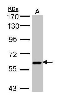
- Sample (30 ug of whole cell lysate)A: H12997.5% SDS PAGEGTX103592 diluted at 1:3000
Supportive validation
- Submitted by
- GeneTex (provider)
- Main image
- Experimental details
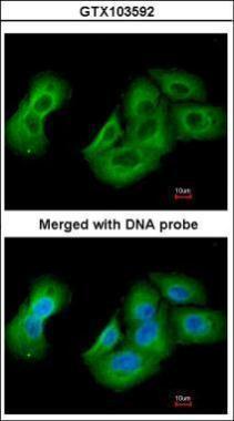
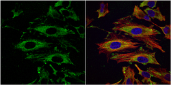
- Immunofluorescence analysis of paraformaldehyde-fixed A549, using UAP1(GTX103592) antibody at 1:200 dilution.
- Submitted by
- GeneTex (provider)
- Main image
- Experimental details
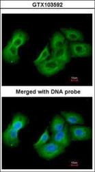
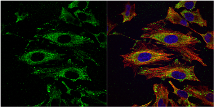
- UAP1 antibody [N1C3] detects UAP1 protein at cytoplasm by immunofluorescent analysis.Sample: HeLa cells were fixed in 4% paraformaldehyde at RT for 15 min.Green: UAP1 protein stained by UAP1 antibody [N1C3] (GTX103592) diluted at 1:200.Red: alpha Tubulin, a cytoskeleton marker, stained by alpha Tubulin antibody [B-5-1-2] (GTX11304) diluted at 1:10000.Blue: Hoechst 33342 staining.
Supportive validation
- Submitted by
- GeneTex (provider)
- Main image

- Experimental details
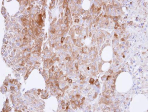
- Immunohistochemical analysis of paraffin-embedded ES-2 xenograft , using UAP1(GTX103592) antibody at 1:500 dilution.