Explore
 Validate
 Learn
LearnSTJ70171
antibody from St John's Laboratory
Targeting: DAP3
bMRP-10, DAP-3, DKFZp686G12159, MGC126058, MGC126059, MRP-S29, MRPS29
 Western blot
 Immunohistochemistry
Antibody data
- Antibody Data
- Antigen structure
- References [0]
- Comments [0]
- Validations
- Immunohistochemistry [1]
- Other assay [3]
Submit
Validation data
Reference
Comment
Report error
- Product number
- STJ70171 - Provider product page

- Provider
- St John's Laboratory
- Product name
- Anti-DAP3 antibody (C-Term) (STJ70171)
- Antibody type
- Polyclonal
- Description
- Goat polyclonal antibody anti-DAP3 (C-Term) is suitable for use in ELISA, Western Blot and Immunohistochemistry research applications.
- Reactivity
- Human
- Host
- Goat
- Conjugate
- Unconjugated
- Antigen sequence
NPSLLERHCAYL- Epitope
- NA
- Isotype
- IgG
- Antibody clone number
- NA
- Vial size
- NA
- Concentration
- NA
- Storage
- Store at-20 on receipt and minimise freeze-thaw cycles.
- Handling
- NA
No comments: Submit comment
Supportive validation
- Submitted by
- St John's Laboratory (provider)
- Main image

- Experimental details
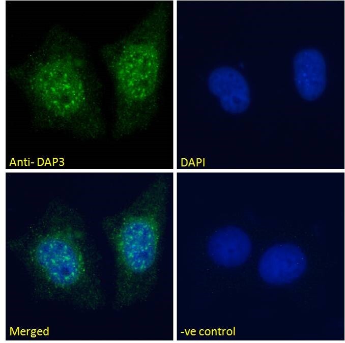
- STJ70171 Immunofluorescence analysis of paraformaldehyde fixed MCF7 cells, permeabilized with 0. 15% Triton. Primary incubation 1hr (10ug/ml) followed by Alexa Fluor 488 secondary antibody (2ug/ml) , showing nuclear staining. The nuclear stain is DAPI (blue). NA NA NA Negative control: Unimmunized goat IgG (10ug/ml) followed by Alexa Fluor 488 secondary antibody (2ug/ml).
- Sample type
- NA
- Validation comment
- NA
- Primary Ab dilution
- NA
- Other comments
- NA
- Secondary Ab
- NA
- Secondary Ab dilution
- NA
- Protocol
- NA
Supportive validation
Supportive validation
Supportive validation
- Submitted by
- St John's Laboratory (provider)
- Main image
- Experimental details
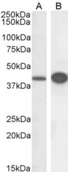
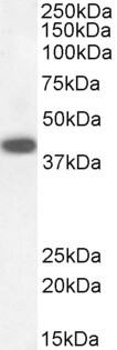
- STJ70171 (0. 3µg/ml) staining of HeLa (A) and HepG2 (B) cell lysate (RIPA buffer, 30µg total protein per lane). Detected by chemiluminescence.
- Sample type
- NA
- Validation comment
- NA
- Primary Ab dilution
- NA
- Other comments
- NA
- Secondary Ab
- NA
- Secondary Ab dilution
- NA
- Protocol
- NA

Supportive validation
- Submitted by
- St John's Laboratory (provider)
- Main image
- Experimental details
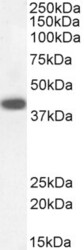
- STJ70171 staining (0. 3µg/ml) of Human Kidney lysate (RIPA buffer, 30µg total protein per lane). Detected by chemiluminescence.
- Sample type
- NA
- Validation comment
- NA
- Primary Ab dilution
- NA
- Other comments
- NA
- Secondary Ab
- NA
- Secondary Ab dilution
- NA
- Protocol
- NA
Supportive validation
- Submitted by
- St John's Laboratory (provider)
- Main image
- Experimental details
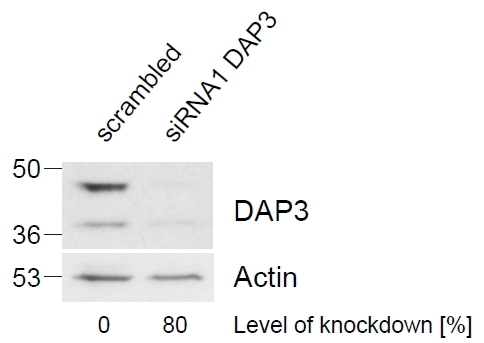
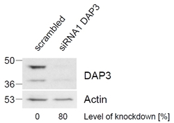
- STJ70171 (1µg/ml) staining of HeLa lysate (control in left lane and after si-RNA-mediated DAP3 knock-down expresson in right lane) (35µg protein in RIPA buffer). Level of knock-down relative to Actin expression level was determined by RT-PCR. Primary incubation was 1 hour. Detected by chemiluminescence. This data is from a previous batch, not on sale.
- Sample type
- NA
- Validation comment
- NA
- Primary Ab dilution
- NA
- Other comments
- NA
- Secondary Ab
- NA
- Secondary Ab dilution
- NA
- Protocol
- NA