 Explore
 Validate
 Learn
Learn Western blot
 Immunocytochemistry
Antibody data
- Antibody Data
- Antigen structure
- References [1]
- Comments [0]
- Validations
- Immunocytochemistry [2]
- Other assay [1]
Submit
Validation data
Reference
Comment
Report error
- Product number
- PA5-66095 - Provider product page

- Provider
- Invitrogen Antibodies
- Product name
- XPOT Polyclonal Antibody
- Antibody type
- Polyclonal
- Antigen
- Recombinant protein fragment
- Description
- Immunogen sequence: MDEQALLGLNP NADSDFRQRA LAYFEQLKIS PDAWQVCAEA LAQRTYSDDH VKFFCFQVLE HQVKYKYSEL TTVQQQLIRE TLISWLQAQM LNPQP Highest antigen sequence identity to the following orthologs - mouse 95%, rat 95%.
- Reactivity
- Human
- Host
- Rabbit
- Isotype
- IgG
- Vial size
- 100 μL
- Concentration
- 0.1 mg/mL
- Storage
- Store at 4°C short term. For long term storage, store at -20°C, avoiding freeze/thaw cycles.
Submitted references Unconventional tonicity-regulated nuclear trafficking of NFAT5 mediated by KPNB1, XPOT and RUVBL2.
Cheung CY, Huang TT, Chow N, Zhang S, Zhao Y, Chau MP, Chan WC, Wong CCL, Boassa D, Phan S, Ellisman MH, Yates JR, Xu S, Yu Z, Zhang Y, Zhang R, Ng LL, Ko BCB
Journal of cell science 2022 Jul 1;135(13)
Journal of cell science 2022 Jul 1;135(13)
No comments: Submit comment
Supportive validation
- Submitted by
- Invitrogen Antibodies (provider)
- Main image
- Experimental details
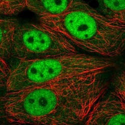
- Immunofluorescent staining of XPOT in human cell line MCF7 shows localization to nucleoplasm and cytosol. Samples were probed using a XPOT Polyclonal Antibody (Product # PA5-66095).

- Submitted by
- Invitrogen Antibodies (provider)
- Main image

- Experimental details
- Immunofluorescent staining of XPOT in human cell line MCF7 shows localization to nucleoplasm and cytosol. Samples were probed using a XPOT Polyclonal Antibody (Product # PA5-66095).

Supportive validation
- Submitted by
- Invitrogen Antibodies (provider)
- Main image
- Experimental details
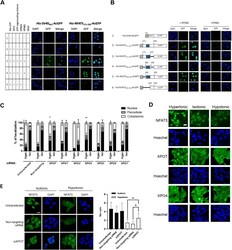
- Fig. 4. Identification of importin and exportin for NFAT5 nuclear import and export. (A) In vitro nuclear import assay of His-SV40 NLS -AcGFP and His-NFAT5 171-250 -AcGFP was carried out using digitonin-permeabilized HeLa cells supplemented with individual nuclear transport factors, as indicated. The transport assay was carried out for 30 min at 37degC. Cells were then fixed with paraformaldehyde and stained with DAPI, and GFP fluorescence images were taken using a confocal microscope. Scale bar: 30 um. Images are representative of three independent experiments. (B) Role of KPNB1 in nuclear import of His-NFAT5-AcGFP. Left: schematic representation of the indicated His-NFAT5-AcGFP reporter constructs. Shaded regions indicate NFAT5-cNLS. Right: in vitro nuclear import assay of His-SV40 NLS -AcGFP and the indicated His-NFAT5-AcGFP fusions was carried out using digitonin-permeabilized HeLa cells supplemented with RanGTP, NTF2 and ATP-regenerating mixture, in the presence or absence of KPNB1. The transport assay was carried out for 30 min at 37degC. Cells were then fixed with paraformaldehyde and stained with DAPI, and images were taken using a confocal microscope. Scale bar: 30 um. Images are representative of three independent experiments. (C) Quantitative analysis of subcellular localization of immunofluorescence signal in HeLa cells transfected with FLAG-NFAT5 132-581 and siRNA targeting the indicated members of the exportin family. Cells were switched to isotonic (Iso) or hyp
