Explore
 Validate
 Learn
Learn Western blot
 ELISA
ELISAAntibody data
- Antibody Data
- Antigen structure
- References [0]
- Comments [0]
- Validations
- Western blot [2]
- Immunohistochemistry [1]
Submit
Validation data
Reference
Comment
Report error
- Product number
- R1555P - Provider product page

- Provider
- Acris Antibodies GmbH
- Proper citation
- Acris Antibodies GmbH Cat#R1555P, RRID:AB_1005587
- Product name
- anti Nestin (1484-1500)
- Antibody type
- Polyclonal
- Antigen
- Recombinant protein corresponding to amino acids 1484-1500 of Human Nestin protein.
- Reactivity
- Human, Mouse
- Host
- Rabbit
- Vial size
- 0.1 mg
- Concentration
- 1.0 mg/ml (by UV absorbance at 280 nm)
No comments: Submit comment
Supportive validation
- Submitted by
- Acris Antibodies GmbH (provider)
- Main image
- Experimental details
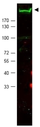
- Western blot using R1555P Nestin antibody shows detection of a band ~220 kDa corresponding to Mouse Nestin (arrowhead). Approximately 30 µg of MEF whole cell lysate was separated by SDS-PAGE using a 4-20% gradient gel. After transfer onto Nitrocellulose, the membrane was blocked and then probed with the primary antibody diluted to 1/2,000 overnight at 4°C. The membrane was then washed and reacted with a 1/10,000 dilution of IRDye(TM)800 conjugated Goatt anti-Rabbit IgG [H&L] for 45 min at RT. IRDye(TM)800 fluorescence image was captured using the Odyssey® Infrared Imaging System developed by LI-COR. IRDye is a trademark of LI-COR, Inc. Other detection systems will yield similar results.

- Submitted by
- Acris Antibodies GmbH (provider)
- Main image
- Experimental details
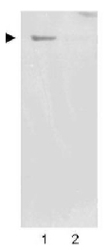
- Western blot using R1555P Nestin antibody shows detection of a band ~220 kDa corresponding to Human Nestin (arrowhead). Undifferentiated HCN-1A Human brain cortical neuron neuronal progenitor lysate (lane 1), or differentiated HCN-1A Human brain cortical neuron neuronal progenitor lysate (lane 2) were separated by SDS-PAGE using 4-20% gradient gel. After transfer onto nitrocellulose, the membrane was blocked and then probed with the primary antibody diluted to 1/2,000 overnight at 4°C. The membrane was then washed and reacted with a 1/10,000 dilution of Peroxidase conjugated Goat anti-Rabbit IgG R1364HRP for 45 min at RT. Image was captured using film. Other detection systems will yield similar results. Image courtesy of Prof. F.H. Gage of the Salk Institute, San Diego, CA.

Supportive validation
- Submitted by
- Acris Antibodies GmbH (provider)
- Main image

- Experimental details
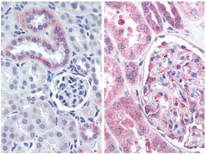
- R1555P Nestin antibody staining of Formalin-Fixed Paraffin-Embedded Mouse Kidney (left) and Human Kidney (Right) at 5 µg/ml