Explore
 Validate
 Learn
Learn Western blot
 ELISA
ELISAAntibody data
- Antibody Data
- Antigen structure
- References [0]
- Comments [0]
- Validations
- Western blot [2]
- ELISA [3]
- Immunohistochemistry [6]
Submit
Validation data
Reference
Comment
Report error
- Product number
- LS-C745287 - Provider product page

- Provider
- LSBio
- Product name
- NES / Nestin Antibody LS-C745287
- Antibody type
- Polyclonal
- Description
- Affinity purified
- Reactivity
- Human, Mouse
- Host
- Rabbit
- Isotype
- IgG
- Storage
- Store vial at -20°C or below prior to opening. Dilute 1:10 to minimize loss. Store the vial at -20°C or below after dilution. Avoid freeze-thaw cycles.
No comments: Submit comment
Enhanced validation
- Submitted by
- LSBio (provider)
- Enhanced method
- Genetic validation
- Main image
- Experimental details
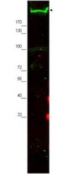
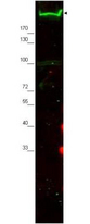
- Western blot using the Affinity Purified anti-Nestin antibody shows detection of a band ~220 kDa corresponding to mouse Nestin (arrowhead). Approximately 30 µg of MEF whole cell lysate was separated by SDS-PAGE using a 4-20% gradient gel. After transfer onto nitrocellulose, the membrane was blocked and then probed with the primary antibody diluted to 1:2,000 overnight at 4°C. The membrane was then washed and reacted with a 1:10,000 dilution of conjugated Gt-a-Rabbit IgG [H&L] MX for 45 min at room temperature. fluorescence image was captured using the Odyssey Infrared Imaging System developed by LI-COR. Other detection systems will yield similar results.
- Submitted by
- LSBio (provider)
- Enhanced method
- Genetic validation
- Main image
- Experimental details
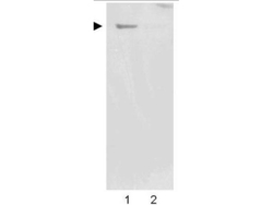
- Western blot using the Affinity Purified anti-Nestin antibody shows detection of a band ~220 kDa corresponding to human Nestin (arrowhead). Undifferentiated HCN-1A human brain cortical neuron neuronal progenitor lysate (lane 1), or differentiated HCN-1A human brain cortical neuron neuronal progenitor lysate (lane 2) were separated by SDS-PAGE using 4-20% gradient gel. After transfer onto nitrocellulose, the membrane was blocked and then probed with the primary antibody diluted to 1:2,000 overnight at 4°C. The membrane was then washed and reacted with a 1:10,000 dilution of peroxidase conjugated affinity purified Gt-a-Rabbit IgG [H&L] MX (611-1302) for 45 min at room temperature. Image was captured using film. Other detection systems will yield similar results.

Supportive validation
- Submitted by
- LSBio (provider)
- Enhanced method
- Genetic validation
- Main image

- Experimental details
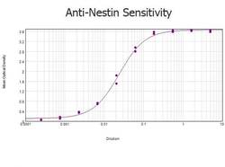
- ELISA results of purified Rabbit anti-Nestin Antibody tested against BSA-conjugated peptide of immunizing peptide. Each well was coated in duplicate with 0.1µg of conjugate. The starting dilution of antibody was 5µg/ml and the X-axis represents the Log10 of a 3-fold dilution. This titration is a 4-parameter curve fit where the IC50 is defined as the titer of the antibody. Assay performed using 3% fish gel, Goat anti-Rabbit IgG Antibody Peroxidase Conjugated (Min X Bv Ch Gt GP Ham Hs Hu Ms Rt & Sh Serum Proteins)

- Submitted by
- LSBio (provider)
- Main image
- Experimental details
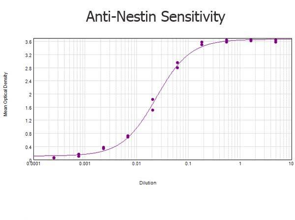
- ELISA results of purified Rabbit anti-Nestin Antibody tested against BSA-conjugated peptide of immunizing peptide. Each well was coated in duplicate with 0.1µg of conjugate. The starting dilution of antibody was 5µg/ml and the X-axis represents the Log10 of a 3-fold dilution. This titration is a 4-parameter curve fit where the IC50 is defined as the titer of the antibody. Assay performed using 3% fish gel, Goat anti-Rabbit IgG Antibody Peroxidase Conjugated (Min X Bv Ch Gt GP Ham Hs Hu Ms Rt & Sh Serum Proteins)
- Submitted by
- LSBio (provider)
- Main image
- Experimental details
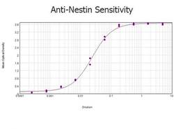
- ELISA results of purified Rabbit anti-Nestin Antibody tested against BSA-conjugated peptide of immunizing peptide. Each well was coated in duplicate with 0.1µg of conjugate. The starting dilution of antibody was 5µg/ml and the X-axis represents the Log10 of a 3-fold dilution. This titration is a 4-parameter curve fit where the IC50 is defined as the titer of the antibody. Assay performed using 3% fish gel, Goat anti-Rabbit IgG Antibody Peroxidase Conjugated (Min X Bv Ch Gt GP Ham Hs Hu Ms Rt & Sh Serum Proteins)

Supportive validation
- Submitted by
- LSBio (provider)
- Enhanced method
- Genetic validation
- Main image
- Experimental details
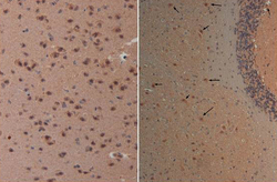
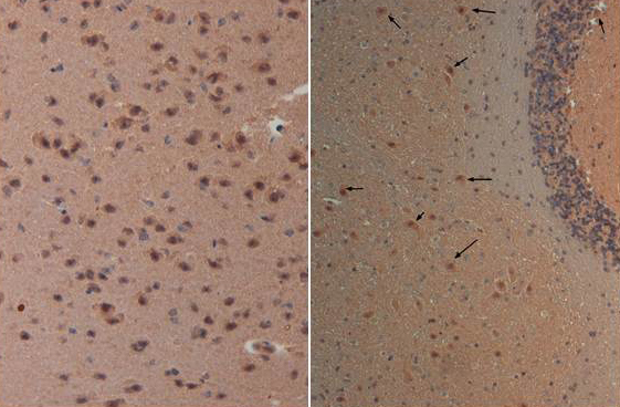
- Immunohistochemistry with Anti-nestin antibody at 40X (left) and 20X (right) Tissue: Brain and Cerebellum (right) Fixation: FFPE buffered formalin 10% conc Antigen retrieval: Heat, Citrate pH 6.2. Pressure Cooker Primary antibody: 20ug/ml 1 hour @ room T Secondary antibody: Goat anti Rabbit Polymer HRP Prediluted by the manufacturer 30 min. @ room T Staining: antibody as precipitated red signal with a hematoxylin purple nuclear counterstain.
- Submitted by
- LSBio (provider)
- Enhanced method
- Genetic validation
- Main image

- Experimental details
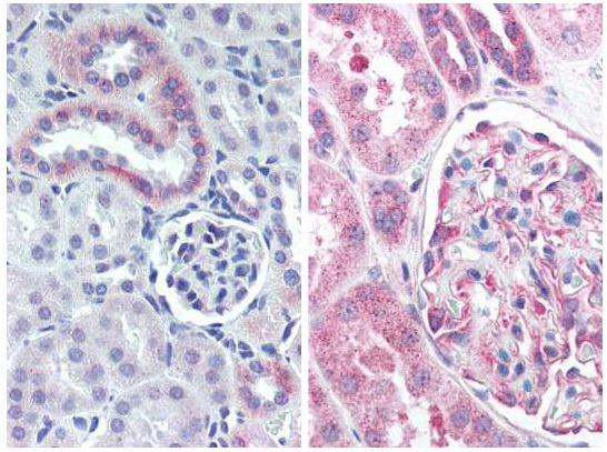
- Immunohistochemistry with Anti-nestin antibody Tissue: mouse kidney (Left) and human kidney (Right). Fixation: formalin-fixed, paraffin-embedded tissue Antigen retrieval: heat-induced Primary antibody: 5 µg/ml Staining: antibody as precipitated red signal with a hematoxylin purple nuclear counterstain.

- Submitted by
- LSBio (provider)
- Enhanced method
- Genetic validation
- Main image

- Experimental details
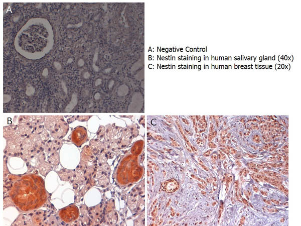
- Immunohistochemistry with anti-nestin antibody showing nestin staining in cytoplasm of of ductal epithelium of human salivary gland (B) and in nucleus and cytoplasm of human breast tissue (C). Formalin fixed/paraffin embedded sections were subjected to heat induced epitope retrieval (HIER) at pH 6.2 and then incubated with rabbit anti-nestin antibody at 4.0 µg/ml for 60 minutes. The reaction was developed using MACH 1 universal HRP polymer detection system and visualized with 3’3-diamino-benzidine substrate (DAB).
- Submitted by
- LSBio (provider)
- Main image

- Experimental details
- Immunohistochemistry with anti-nestin antibody showing nestin staining in cytoplasm of of ductal epithelium of human salivary gland (B) and in nucleus and cytoplasm of human breast tissue (C). Formalin fixed/paraffin embedded sections were subjected to heat induced epitope retrieval (HIER) at pH 6.2 and then incubated with rabbit anti-nestin antibody at 4.0 µg/ml for 60 minutes. The reaction was developed using MACH 1 universal HRP polymer detection system and visualized with 3’3-diamino-benzidine substrate (DAB).

- Submitted by
- LSBio (provider)
- Main image

- Experimental details
- Immunohistochemistry with Anti-nestin antibody at 40X (left) and 20X (right) Tissue: Brain and Cerebellum (right) Fixation: FFPE buffered formalin 10% conc Antigen retrieval: Heat, Citrate pH 6.2. Pressure Cooker Primary antibody: 20ug/ml 1 hour @ room T Secondary antibody: Goat anti Rabbit Polymer HRP Prediluted by the manufacturer 30 min. @ room T Staining: antibody as precipitated red signal with a hematoxylin purple nuclear counterstain.

- Submitted by
- LSBio (provider)
- Main image

- Experimental details
- Immunohistochemistry with Anti-nestin antibody Tissue: mouse kidney (Left) and human kidney (Right). Fixation: formalin-fixed, paraffin-embedded tissue Antigen retrieval: heat-induced Primary antibody: 5 µg/ml Staining: antibody as precipitated red signal with a hematoxylin purple nuclear counterstain.