Explore
 Validate
 Learn
Learn Western blot
 Immunocytochemistry
Antibody data
- Antibody Data
- Antigen structure
- References [20]
- Comments [0]
- Validations
- Western blot [1]
- Immunohistochemistry [2]
- Flow cytometry [5]
Submit
Validation data
Reference
Comment
Report error
- Product number
- NB300-266 - Provider product page

- Provider
- Novus Biologicals
- Proper citation
- Novus Cat#NB300-266, RRID:AB_10001441
- Product name
- Mouse Monoclonal Nestin Antibody
- Antibody type
- Monoclonal
- Description
- Protein G purified.
- Reactivity
- Human, Mouse, Rat, Rabbit
- Host
- Mouse
- Isotype
- IgG
- Vial size
- 0.1 ml
- Concentration
- 2.4 mg/ml
- Storage
- Store at 4C short term. Aliquot and store at -20C long term. Avoid freeze-thaw cycles.
Submitted references Changes in vitreal protein profile and retina mRNAs in Reeler mice: NGF, IL33 and Müller cell activation.
An increased skin microvessel density is associated with neurovascular complications in type 1 diabetes mellitus.
Dermal microvessel density and maturity is closely associated with atherogenic dyslipidemia and accumulation of advanced glycation end products in adult patients with type 1 diabetes.
Tyrosinase and nestin immunohistochemical expression in melanocytic nevi as a histopathologic pattern to trace melanocyte differentiation and nevogenesis.
Driving Neuronal Differentiation through Reversal of an ERK1/2-miR-124-SOX9 Axis Abrogates Glioblastoma Aggressiveness.
Immunophenotypic characterization of telocyte-like cells in pterygium.
Establishment of a tumor sphere cell line from a metastatic brain neuroendocrine tumor.
Activated Notch1 expression is associated with angiogenesis in cutaneous melanoma.
Comprehensive characterization of glioblastoma tumor tissues for biomarker identification using mass spectrometry-based label-free quantitative proteomics.
Nestin and vimentin colocalization affects the subcellular location of glucocorticoid receptor in cutaneous melanoma.
Epstein-Barr virus latent membrane protein 1 induces cancer stem/progenitor-like cells in nasopharyngeal epithelial cell lines.
Illumina whole-genome complementary DNA-mediated annealing, selection, extension and ligation platform: assessing its performance in formalin-fixed, paraffin-embedded samples and identifying invasion pattern-related genes in oral squamous cell carcinoma.
Comparative analysis of DNA repair in stem and nonstem glioma cell cultures.
NK cells recognize and kill human glioblastoma cells with stem cell-like properties.
SOX2 silencing in glioblastoma tumor-initiating cells causes stop of proliferation and loss of tumorigenicity.
Differential processing of amyloid-beta precursor protein directs human embryonic stem cell proliferation and differentiation into neuronal precursor cells.
Gene expression in human neural stem cells: effects of leukemia inhibitory factor.
Gene expression in human neural stem cells: effects of leukemia inhibitory factor.
Analysis of the temporal expression of nestin in human fetal brain derived neuronal and glial progenitor cells.
Analysis of the temporal expression of nestin in human fetal brain derived neuronal and glial progenitor cells.
Balzamino BO, Esposito G, Marino R, Keller F, Micera A
PloS one 2019;14(2):e0212732
PloS one 2019;14(2):e0212732
An increased skin microvessel density is associated with neurovascular complications in type 1 diabetes mellitus.
Adamska A, Pilacinski S, Zozulinska-Ziolkiewicz D, Gandecka A, Grzelka A, Konwerska A, Malinska A, Nowicki M, Araszkiewicz A
Diabetes & vascular disease research 2019 Nov;16(6):513-522
Diabetes & vascular disease research 2019 Nov;16(6):513-522
Dermal microvessel density and maturity is closely associated with atherogenic dyslipidemia and accumulation of advanced glycation end products in adult patients with type 1 diabetes.
Adamska A, Araszkiewicz A, Pilacinski S, Gandecka A, Grzelka A, Kowalska K, Malinska A, Nowicki M, Zozulinska-Ziolkiewicz D
Microvascular research 2019 Jan;121:46-51
Microvascular research 2019 Jan;121:46-51
Tyrosinase and nestin immunohistochemical expression in melanocytic nevi as a histopathologic pattern to trace melanocyte differentiation and nevogenesis.
Murtas D, Pilloni L, Diana A, Casula L, Tomei S, Piras F, Ferreli C, Maxia C, Perra MT
Histochemistry and cell biology 2019 Feb;151(2):175-185
Histochemistry and cell biology 2019 Feb;151(2):175-185
Driving Neuronal Differentiation through Reversal of an ERK1/2-miR-124-SOX9 Axis Abrogates Glioblastoma Aggressiveness.
Sabelström H, Petri R, Shchors K, Jandial R, Schmidt C, Sacheva R, Masic S, Yuan E, Fenster T, Martinez M, Saxena S, Nicolaides TP, Ilkhanizadeh S, Berger MS, Snyder EY, Weiss WA, Jakobsson J, Persson AI
Cell reports 2019 Aug 20;28(8):2064-2079.e11
Cell reports 2019 Aug 20;28(8):2064-2079.e11
Immunophenotypic characterization of telocyte-like cells in pterygium.
Maxia C, Murtas D, Isola M, Tamma R, Zucca I, Piras F, Ribatti D, Diana A, Perra MT
Molecular vision 2018;24:853-866
Molecular vision 2018;24:853-866
Establishment of a tumor sphere cell line from a metastatic brain neuroendocrine tumor.
Iwata R, Maruyama M, Ito T, Nakano Y, Kanemura Y, Koike T, Oe S, Yoshimura K, Nonaka M, Nomura S, Sugimoto T, Yamada H, Asai A
Medical molecular morphology 2017 Dec;50(4):211-219
Medical molecular morphology 2017 Dec;50(4):211-219
Activated Notch1 expression is associated with angiogenesis in cutaneous melanoma.
Murtas D, Piras F, Minerba L, Maxia C, Ferreli C, Demurtas P, Lai S, Mura E, Corrias M, Sirigu P, Perra MT
Clinical and experimental medicine 2015 Aug;15(3):351-60
Clinical and experimental medicine 2015 Aug;15(3):351-60
Comprehensive characterization of glioblastoma tumor tissues for biomarker identification using mass spectrometry-based label-free quantitative proteomics.
Heroux MS, Chesnik MA, Halligan BD, Al-Gizawiy M, Connelly JM, Mueller WM, Rand SD, Cochran EJ, LaViolette PS, Malkin MG, Schmainda KM, Mirza SP
Physiological genomics 2014 Jul 1;46(13):467-81
Physiological genomics 2014 Jul 1;46(13):467-81
Nestin and vimentin colocalization affects the subcellular location of glucocorticoid receptor in cutaneous melanoma.
Lai S, Piras F, Spiga S, Perra MT, Minerba L, Piga M, Mura E, Murtas D, Demurtas P, Corrias M, Maxia C, Ferreli C, Sirigu P
Histopathology 2013 Feb;62(3):487-98
Histopathology 2013 Feb;62(3):487-98
Epstein-Barr virus latent membrane protein 1 induces cancer stem/progenitor-like cells in nasopharyngeal epithelial cell lines.
Kondo S, Wakisaka N, Muramatsu M, Zen Y, Endo K, Murono S, Sugimoto H, Yamaoka S, Pagano JS, Yoshizaki T
Journal of virology 2011 Nov;85(21):11255-64
Journal of virology 2011 Nov;85(21):11255-64
Illumina whole-genome complementary DNA-mediated annealing, selection, extension and ligation platform: assessing its performance in formalin-fixed, paraffin-embedded samples and identifying invasion pattern-related genes in oral squamous cell carcinoma.
Loudig O, Brandwein-Gensler M, Kim RS, Lin J, Isayeva T, Liu C, Segall JE, Kenny PA, Prystowsky MB
Human pathology 2011 Dec;42(12):1911-22
Human pathology 2011 Dec;42(12):1911-22
Comparative analysis of DNA repair in stem and nonstem glioma cell cultures.
Ropolo M, Daga A, Griffero F, Foresta M, Casartelli G, Zunino A, Poggi A, Cappelli E, Zona G, Spaziante R, Corte G, Frosina G
Molecular cancer research : MCR 2009 Mar;7(3):383-92
Molecular cancer research : MCR 2009 Mar;7(3):383-92
NK cells recognize and kill human glioblastoma cells with stem cell-like properties.
Castriconi R, Daga A, Dondero A, Zona G, Poliani PL, Melotti A, Griffero F, Marubbi D, Spaziante R, Bellora F, Moretta L, Moretta A, Corte G, Bottino C
Journal of immunology (Baltimore, Md. : 1950) 2009 Mar 15;182(6):3530-9
Journal of immunology (Baltimore, Md. : 1950) 2009 Mar 15;182(6):3530-9
SOX2 silencing in glioblastoma tumor-initiating cells causes stop of proliferation and loss of tumorigenicity.
Gangemi RM, Griffero F, Marubbi D, Perera M, Capra MC, Malatesta P, Ravetti GL, Zona GL, Daga A, Corte G
Stem cells (Dayton, Ohio) 2009 Jan;27(1):40-8
Stem cells (Dayton, Ohio) 2009 Jan;27(1):40-8
Differential processing of amyloid-beta precursor protein directs human embryonic stem cell proliferation and differentiation into neuronal precursor cells.
Porayette P, Gallego MJ, Kaltcheva MM, Bowen RL, Vadakkadath Meethal S, Atwood CS
The Journal of biological chemistry 2009 Aug 28;284(35):23806-17
The Journal of biological chemistry 2009 Aug 28;284(35):23806-17
Gene expression in human neural stem cells: effects of leukemia inhibitory factor.
Wright LS, Li J, Caldwell MA, Wallace K, Johnson JA, Svendsen CN
Journal of neurochemistry 2003 Jul;86(1):179-95
Journal of neurochemistry 2003 Jul;86(1):179-95
Gene expression in human neural stem cells: effects of leukemia inhibitory factor.
Wright LS, Li J, Caldwell MA, Wallace K, Johnson JA, Svendsen CN
Journal of neurochemistry 2003 Jul;86(1):179-95
Journal of neurochemistry 2003 Jul;86(1):179-95
Analysis of the temporal expression of nestin in human fetal brain derived neuronal and glial progenitor cells.
Messam CA, Hou J, Berman JW, Major EO
Brain research. Developmental brain research 2002 Mar 31;134(1-2):87-92
Brain research. Developmental brain research 2002 Mar 31;134(1-2):87-92
Analysis of the temporal expression of nestin in human fetal brain derived neuronal and glial progenitor cells.
Messam CA, Hou J, Berman JW, Major EO
Brain research. Developmental brain research 2002 Mar 31;134(1-2):87-92
Brain research. Developmental brain research 2002 Mar 31;134(1-2):87-92
No comments: Submit comment
Supportive validation
- Submitted by
- Novus Biologicals (provider)
- Main image
- Experimental details
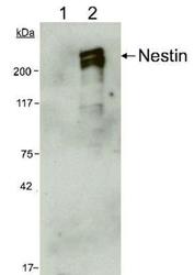
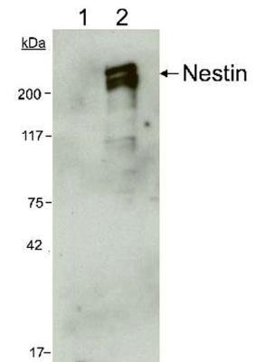
- Western Blot: Nestin Antibody (10C2) [NB300-266] - WB analysis of 5 ug lysate from (1) Rat's total brain tissue, and (2) human CNS progenitor cells using Nestin antibody (clone 10C2) at 1:1000 dilution. The antibody did not react with rat, whereas, in lysate from human samples, it generated a doublet band representing the phosphorylated and the non-phosphorylated forms of Nestin protein.
Supportive validation
- Submitted by
- Novus Biologicals (provider)
- Main image
- Experimental details
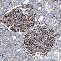
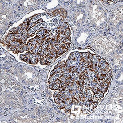
- Immunohistochemistry-Paraffin: Nestin Antibody (10C2) [NB300-266] - Nestin was detected in immersion fixed paraffin-embedded sections of human kidney using anti-human mouse monoclonal antibody (Catalog # NB300-266) at 1:500 dilution overnight at 4C. Tissue was stained using the VisuCyte anti-mouse HRP polymer detection reagent (Catalog # VC001) with DAB chromogen (brown) and counterstained with hematoxylin (blue).Images may not be copied, printed or otherwise disseminated without express written permission of Novus Biologicals a bio-techne brand.
- Submitted by
- Novus Biologicals (provider)
- Main image
- Experimental details
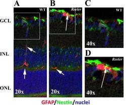
- Immunohistochemistry: Nestin Antibody (10C2) [NB300-266] - GFAP and Nestin immunoreactivity in retinal sections. Epifluorescent acquisition of p28 Reeler and WT retinas. (A-D) As compared to WT, both GFAP (red) and Nestin (green) immunoreactivities were highly visible in Reeler retinas (AB, GCL). Arrows point at yellow immunoreactivity in the white frame (B) indicating GFAP and Nestin co-expression in cells having long-filaments (activated Muller cells), as compared to wild type (arrowheads in A). Nuclei were DAPI counterstained (blue). Image collected and cropped by CiteAb from the following publication (//doi.org/10.1371/journal.pone.0212732) licensed under a CC-BY licence.

Supportive validation
- Submitted by
- Novus Biologicals (provider)
- Main image
- Experimental details
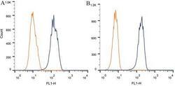
- Flow Cytometry: Nestin Antibody (10C2) [NB300-266] - Intracellular flow cytometric staining of 1 x 10^6 CHO (A) and HEK-293 (B) cells using Nestin antibody (dark blue). Isotype control shown in orange. An antibody concentration of 1 ug/1 x 10^6 cells was used.
- Submitted by
- Novus Biologicals (provider)
- Main image

- Experimental details
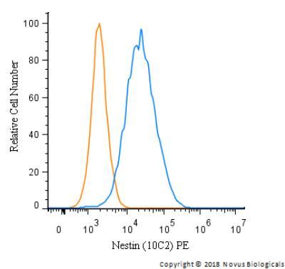
- Flow Cytometry: Nestin Antibody (10C2) [NB300-266] - An intracellular stain was performed on HeLa cells with Nestin Antibody (10C2)NB300-266PE (blue) and a matched isotype control (orange). Cells were fixed with 4% PFA and then permeabilized with 0.1% saponin. Cells were incubated in an antibody dilution of 2.5 ug/mL for 30 minutes at room temperature. Both antibodies were conjugated to phycoerythrin.
- Submitted by
- Novus Biologicals (provider)
- Main image
- Experimental details
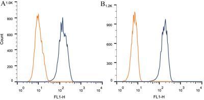
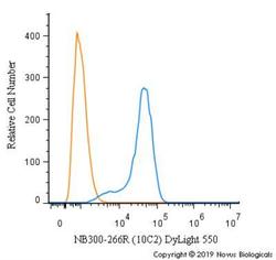
- Flow Cytometry: Nestin Antibody (10C2) [NB300-266] - An intracellular stain was performed on SH-SY5Y cells with Nestin [10C2] Antibody NB300-266R (blue) and a matched isotype control (orange). Cells were fixed with 4% PFA and then permeabilized with 0.1% saponin. Cells were incubated in an antibody dilution of 10 ug/mL for 30 minutes at room temperature. Both antibodies were conjugated to DyLight 550.
- Submitted by
- Novus Biologicals (provider)
- Main image
- Experimental details
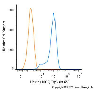
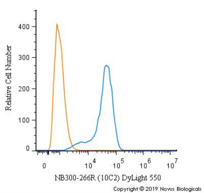
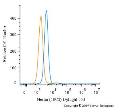
- Flow Cytometry: Nestin Antibody (10C2) [NB300-266] - An intracellular stain was performed on SK-MEL-28 cells with Nestin [10C2] Antibody NB300-266R (blue) and a matched isotype control (orange). Cells were fixed with 4% PFA and then permeabilized with 0.1% saponin. Cells were incubated in an antibody dilution of 5 ug/mL for 30 minutes at room temperature. Both antibodies were conjugated to DyLight 550.
- Submitted by
- Novus Biologicals (provider)
- Main image
- Experimental details
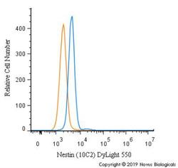
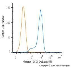
- Flow Cytometry: Nestin Antibody (10C2) [NB300-266] - An intracellular stain was performed on SH-SY5Y cells with Nestin [10C2] Antibody NB300-266C (blue) and a matched isotype control (orange). Cells were fixed with 4% PFA and then permeabilized with 0.1% saponin. Cells were incubated in an antibody dilution of 2.5 ug/mL for 30 minutes at room temperature. Both antibodies were conjugated to DyLight 650.