 Explore
 Validate
 Learn
Learn Western blot
Antibody data
- Antibody Data
- Antigen structure
- References [0]
- Comments [0]
- Validations
- Western blot [9]
- Immunocytochemistry [2]
- Immunohistochemistry [1]
Submit
Validation data
Reference
Comment
Report error
- Product number
- MA1-20344 - Provider product page

- Provider
- Invitrogen Antibodies
- Product name
- TBK1 Monoclonal Antibody (108A429)
- Antibody type
- Monoclonal
- Antigen
- Synthetic peptide
- Description
- MA1-20344 detects NAK in human samples.
- Reactivity
- Human, Mouse, Rat, Bovine, Canine
- Host
- Mouse
- Isotype
- IgG
- Antibody clone number
- 108A429
- Vial size
- 50 µg
- Concentration
- 1.0 mg/mL
- Storage
- Store at 4°C short term. For long term storage, store at -20°C, avoiding freeze/thaw cycles.
No comments: Submit comment
Supportive validation
- Submitted by
- Invitrogen Antibodies (provider)
- Main image

- Experimental details
- Western blot of NAK in lysate from 293 cells untransfected (lane 1) and transfected with human TBK1 cDNA (lane 2) using a NAK monoclonal antibody (Product # MA1-20344) at a dilution of 2 µg/mL.

- Submitted by
- Invitrogen Antibodies (provider)
- Main image

- Experimental details
- Western blot analysis of NAK in Daudi (A) and mouse RAW (B) cell lysate using a NAK monoclonal antibody (Product # MA1-20344) at a dilution of 2 µg/mL.

- Submitted by
- Invitrogen Antibodies (provider)
- Main image

- Experimental details
- Western Blot analysis of TBK1 was performed by loading Daudi (left) and Raw 264.7 (right) cell lysates. Proteins were transferred to a membrane and probed with a TBK1 Monoclonal Antibody (108A429) (Product # MA1-20344).

- Submitted by
- Invitrogen Antibodies (provider)
- Main image

- Experimental details
- Western Blot analysis of TBK1 was performed by loading Daudi (A) and RAW264.7 (B) cell lysates. Proteins were transferred to a membrane and probed with a TBK1 Monoclonal Antibody (108A429) (Product # MA1-20344) at a dilution of 2 µg/mL.

- Submitted by
- Invitrogen Antibodies (provider)
- Main image

- Experimental details
- Western blot was performed using Anti-TBK1 Monoclonal Antibody (108A429) (Product # MA1-20344) and a 83 kDa band corresponding to TBK1 was observed across cell lines and tissues tested. Whole cell extracts (30 µg lysate) of HEK-293 (Lane 1), HeLa (Lane 2), K-562 (Lane 3), MCF7 (Lane 4), T-47D (Lane 5) and SK-BR-3 (Lane 6), tissue extracts of Mouse Heart (Lane 8), Rat Heart (Lane 7), Mouse Testis (Lane 9) and Rat Testis (Lane 10) were electrophoresed using NuPAGE™ 4-12% Bis-Tris Protein Gel (Product # NP0322BOX). Resolved proteins were then transferred onto a nitrocellulose membrane (Product # IB23002) by iBlot® 2 Dry Blotting System (Product # IB21001) and then blocked with Pierce™ Protein-Free T20 (PBS) Blocking Buffe (Product # 37573). The blot was probed with the primary antibody (1 µg/mL) and detected by chemiluminescence with Goat anti-Mouse IgG (H+L) Superclonal™ Recombinant Secondary Antibody, HRP (Product # A28177, 1:20000 dilution) using the iBright™ FL1500 Imaging System (Product # A44115). Chemiluminescent detection was performed using SuperSignal™ West Pico PLUS Chemiluminescent Substrate (Product # 34580).

- Submitted by
- Invitrogen Antibodies (provider)
- Main image

- Experimental details
- Knockout of TBK1 was achieved by CRISPR-Cas9 genome editing using LentiArray™ Lentiviral sgRNA (Product # A32042, Assay ID CRISPR1012237_LV) and LentiArray Cas9 Lentivirus (Product # A32064). Western blot analysis of TBK1 was performed by loading 30 µg of A549 wild type (Lane 1), A549 Cas9 (Lane 2) andA549 TBK1 KO (Lane 3) whole cell extracts. The samples were electrophoresed using NuPAGE™ Novex™ 4-12% Bis-Tris Protein Gel (Product # NP0322BOX). Resolved proteins were then transferred onto a nitrocellulose membrane (Product # IB23001) by iBlot® 2 Dry Blotting System (Product # IB21001). The blot was probed with TBK1 Monoclonal Antibody (108A429) (Product # MA1-20344, 1:500 dilution) and Goat anti-Mouse IgG (H+L) Superclonal™ Recombinant Secondary Antibody, HRP (Product # A28177, 1:10,000 dilution) using the iBright™ FL 1500 (Product # A44115). Chemiluminescent detection was performed using SuperSignal™ West Atto Ultimate Sensitivity Substrate (Product # A38556). Loss of signal upon CRISPR mediated knockout (KO) using the LentiArray™ CRISPR product line confirms that antibody is specific to TBK1.

- Submitted by
- Invitrogen Antibodies (provider)
- Main image

- Experimental details
- Western Blot analysis of TBK1 was performed by loading Daudi (left) and Raw 264.7 (right) cell lysates. Proteins were transferred to a membrane and probed with a TBK1 Monoclonal Antibody (108A429) (Product # MA1-20344).

- Submitted by
- Invitrogen Antibodies (provider)
- Main image

- Experimental details
- Knockdown of TBK1 was achieved by transfecting HEK-293 with TBK1 specific siRNAs (Silencer® select Product # S763). Western blot analysis (Fig. a) was performed using Whole cell extracts from the TBK1 knockdown cells (lane 3), non-targeting scrambled siRNA transfected cells (lane 2) and untransfected cells (lane 1). The blot was probed with TBK1 Monoclonal Antibody (108A429) (Product # MA1-20344, 1 µg/mL ) and Goat anti-Mouse IgG (H+L) Superclonal™ Recombinant Secondary Antibody, HRP (Product # A28177, 1:20000 dilution). Densitometric analysis of this western blot is shown in histogram (Fig. b). Decrease in signal upon siRNA mediated knock down confirms that antibody is specific to TBK1.

- Submitted by
- Invitrogen Antibodies (provider)
- Main image

- Experimental details
- Western Blot analysis of TBK1 was performed by loading HEK293 mock cells (Lane 1) or overexpressing human NAK. Proteins were transferred to a membrane and probed with a TBK1 Monoclonal Antibody (108A429) (Product # MA1-20344) at a dilution of 2 µg/mL.

Supportive validation
- Submitted by
- Invitrogen Antibodies (provider)
- Main image
- Experimental details
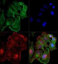
- Immunocytochemistry-Immunofluorescence analysis of TBK1 in HeLa cells using TBK1 Monoclonal Antibody (108A429) (Product # MA1-20344) (Green) at a dilution of 1:10. Red : Tubulin. Blue: DAPI.

- Submitted by
- Invitrogen Antibodies (provider)
- Main image
- Experimental details
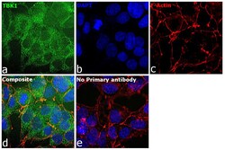
- Immunofluorescence analysis of TBK1 was performed using 70% confluent log phase HEK-293 cells. The cells were fixed with 4% paraformaldehyde for 10 minutes, permeabilized with 0.1% Triton™ X-100 for 15 minutes, and blocked with 2% BSA for 45 minutes at room temperature. The cells were labeled with TBK1 Monoclonal Antibody (108A429) (Product # MA1-20344) at 1:100 in 0.1% BSA, incubated at 4 degree celsius overnight and then labeled with Donkey anti-Mouse IgG (H+L) Highly Cross-Adsorbed Secondary Antibody, Alexa Fluor Plus 488 (Product # A32766), (1:2000), for 45 minutes at room temperature (Panel a: Green). Nuclei (Panel b:Blue) were stained with Hoechst 33342 (Product # H1399). F-actin (Panel c: Red) was stained with Alexa Fluor™ Plus 647 Phalloidin (Product # A30107, 1:2000 dilution). Panel d represents the merged image showing cytoplasmic localization. Panel e represents control cells with no primary antibody to assess background. The images were captured at 40 magnification.

Supportive validation
- Submitted by
- Invitrogen Antibodies (provider)
- Main image
- Experimental details
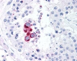
- Immunohistochemistry (Paraffin) analysis of TBK1 in human testis tissue using TBK1 Monoclonal Antibody (108A429) (Product # MA1-20344).
