Explore
 Validate
 Learn
Learn Western blot
Antibody data
- Antibody Data
- Antigen structure
- References [0]
- Comments [0]
- Validations
- Western blot [4]
- Immunocytochemistry [1]
- Immunohistochemistry [2]
Submit
Validation data
Reference
Comment
Report error
- Product number
- PA5-34809 - Provider product page

- Provider
- Invitrogen Antibodies
- Product name
- TBK1 Polyclonal Antibody
- Antibody type
- Polyclonal
- Antigen
- Recombinant protein fragment
- Description
- Application Note: For IHC, epitope retrieval with citrate buffer pH 6.0 is recommended for FFPE tissue sections.
- Reactivity
- Human, Mouse
- Host
- Rabbit
- Isotype
- IgG
- Vial size
- 100 µL
- Concentration
- 0.7 mg/mL
- Storage
- Store at 4°C short term. For long term storage, store at -20°C, avoiding freeze/thaw cycles.
No comments: Submit comment
Supportive validation
- Submitted by
- Invitrogen Antibodies (provider)
- Main image
- Experimental details
- Western Blot analysis of TBK1 was performed by separating 30 µg of whole cell extracts by 7.5% SDS-PAGE. Proteins were transferred to a membrane and probed with a TBK1 Polyclonal Antibody (Product # PA5-34809) at a dilution of 1:1000.
- Submitted by
- Invitrogen Antibodies (provider)
- Main image
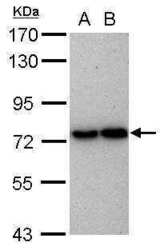
- Experimental details
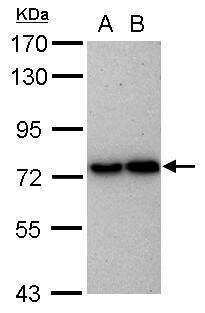
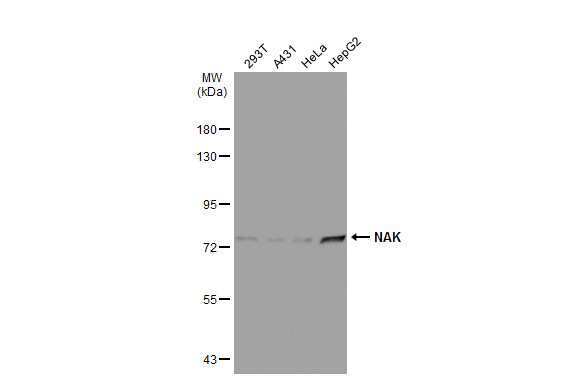
- Western Blot using TBK1 Polyclonal Antibody (Product # PA5-34809). Sample (30 µg of whole cell lysate). Lane A: HeLa. Lane B: HepG2 . 7.5% SDS PAGE. TBK1 Polyclonal Antibody (Product # PA5-34809) diluted at 1:1,000.
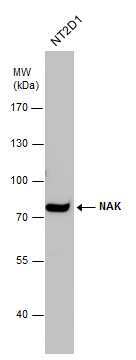
- Submitted by
- Invitrogen Antibodies (provider)
- Main image
- Experimental details
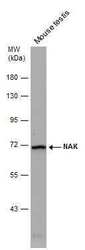
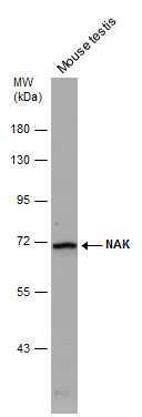
- Western Blot analysis of TBK1 was performed by separating 50 µg of Mouse tissue extracts by 7.5% SDS-PAGE. Proteins were transferred to a membrane and probed with a TBK1 Polyclonal Antibody (Product # PA5-34809) at a dilution of 1:500.
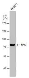
- Submitted by
- Invitrogen Antibodies (provider)
- Main image
- Experimental details
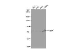
- Western Blot analysis of TBK1 was performed by separating 30 µg of various whole cell extracts by 7.5% SDS-PAGE. Proteins were transferred to a membrane and probed with a TBK1 Polyclonal Antibody (Product # PA5-34809) at a dilution of 1:1000 and a HRP-conjugated anti-rabbit IgG secondary antibody.
Supportive validation
- Submitted by
- Invitrogen Antibodies (provider)
- Main image

- Experimental details
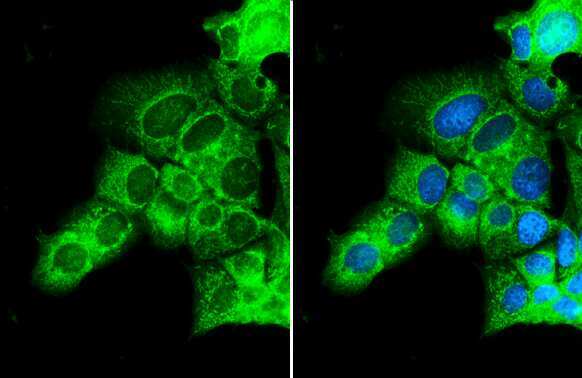
- NAK/TBK1 antibody [N2C1], Internal detects NAK/TBK1 protein at cytoplasm by immunofluorescent analysis. Sample: MCF-7 cells were fixed in 4% paraformaldehyde at RT for 15 min. Green: NAK/TBK1 stained by NAK/TBK1 antibody [N2C1], Internal (Product # PA5-34809) diluted at 1:500. Blue: Fluoroshield with DAPI .
Supportive validation
- Submitted by
- Invitrogen Antibodies (provider)
- Main image
- Experimental details
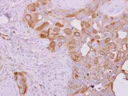
- NAK/TBK1 antibody [N2C1], Internal detects NAK/TBK1 protein at cytoplasm on H441 xenograft by immunohistochemical analysis. Sample: Paraffin-embedded H441 xenograft. NAK/TBK1 antibody [N2C1], Internal (Product # PA5-34809) dilution: 1:500. Antigen Retrieval: EDTA based, pH 8.0 buffer, 15 min.

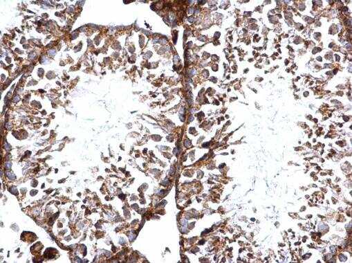
- Submitted by
- Invitrogen Antibodies (provider)
- Main image
- Experimental details
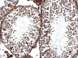
- NAK/TBK1 antibody [N2C1], Internal detects NAK/TBK1 protein at cytosol and nucleus on mouse testis by immunohistochemical analysis. Sample: Paraffin-embedded mouse testis. NAK/TBK1 antibody [N2C1], Internal (Product # PA5-34809) dilution: 1:500. Antigen Retrieval: EDTA based, pH 8.0 buffer, 15 min.