Explore
 Validate
 Learn
Learn Western blot
 Immunocytochemistry
Antibody data
- Antibody Data
- Antigen structure
- References [1]
- Comments [0]
- Validations
- Immunocytochemistry [1]
Submit
Validation data
Reference
Comment
Report error
- Product number
- 703154 - Provider product page

- Provider
- Invitrogen Antibodies
- Product name
- TBK1 Recombinant Rabbit Monoclonal Antibody (12H60L39)
- Antibody type
- Monoclonal
- Antigen
- Synthetic peptide
- Reactivity
- Human, Mouse
- Host
- Rabbit
- Isotype
- IgG
- Antibody clone number
- 12H60L39
- Vial size
- 100 µg
- Concentration
- 0.5 mg/mL
- Storage
- Store at 4°C short term. For long term storage, store at -20°C, avoiding freeze/thaw cycles.
Submitted references The A137R Protein of African Swine Fever Virus Inhibits Type I Interferon Production via the Autophagy-Mediated Lysosomal Degradation of TBK1.
Sun M, Yu S, Ge H, Wang T, Li Y, Zhou P, Pan L, Han Y, Yang Y, Sun Y, Li S, Li LF, Qiu HJ
Journal of virology 2022 May 11;96(9):e0195721
Journal of virology 2022 May 11;96(9):e0195721
No comments: Submit comment
Supportive validation
- Submitted by
- Invitrogen Antibodies (provider)
- Main image
- Experimental details
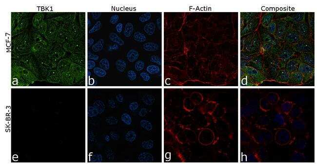
- For immunofluorescence analysis, MCF-7 and SK-BR-3 cells were fixed and permeabilized for detection of endogenous TBK1 using Anti-TBK1 Recombinant Rabbit Monoclonal Antibody (Product # 703154, 1:100) and labeled with Goat anti-Rabbit IgG (H+L) Highly Cross-Adsorbed Secondary Antibody, Alexa Fluor Plus 488 conjugate (Product # A32731, 1:2000). Nuclei (blue) were stained using ProLong™ Diamond Antifade Mountant with DAPI (Product # P36962), and Rhodamine Phalloidin (Product # R415, 1:800) was used for cytoskeletal F-actin (red) staining. Panel a-d) clearly demonstrates the nuclear and cytoplasmic localization of TBK1 in MDA-MB-231 cells. Panel e-h) shows reduced specific staining in SK-BR-3 cells which has low expression compared to MDA-MB-231, demonstrating specificity. The images were captured at 60X magnification.