Antibody data
- Antibody Data
- Antigen structure
- References [2]
- Comments [0]
- Validations
- Western blot [5]
- Immunocytochemistry [1]
- Other assay [1]
Submit
Validation data
Reference
Comment
Report error
- Product number
- PA5-17478 - Provider product page
- Provider
- Invitrogen Antibodies
- Product name
- TBK1 Polyclonal Antibody
- Antibody type
- Polyclonal
- Antigen
- Synthetic peptide
- Description
- It is not recommended to aliquot this antibody.
- Reactivity
- Human, Mouse, Rat
- Host
- Rabbit
- Isotype
- IgG
- Vial size
- 100 µL
- Concentration
- 174 µg/mL
- Storage
- -20°C
Submitted references Association of the TBK1 mutation p.Ile334Thr with frontotemporal dementia and literature review.
Haploinsufficiency of TBK1 causes familial ALS and fronto-temporal dementia.
Yu H, Yu W, Luo SS, Yang YJ, Liu FT, Zhang Y, Chen Y, Sun YM, Wu JJ
Molecular genetics & genomic medicine 2019 Mar;7(3):e547
Haploinsufficiency of TBK1 causes familial ALS and fronto-temporal dementia.
Freischmidt A, Wieland T, Richter B, Ruf W, Schaeffer V, Müller K, Marroquin N, Nordin F, Hübers A, Weydt P, Pinto S, Press R, Millecamps S, Molko N, Bernard E, Desnuelle C, Soriani MH, Dorst J, Graf E, Nordström U, Feiler MS, Putz S, Boeckers TM, Meyer T, Winkler AS, Winkelman J, de Carvalho M, Thal DR, Otto M, Brännström T, Volk AE, Kursula P, Danzer KM, Lichtner P, Dikic I, Meitinger T, Ludolph AC, Strom TM, Andersen PM, Weishaupt JH
Nature neuroscience 2015 May;18(5):631-6
No comments: Submit comment
Supportive validation
- Submitted by
- Invitrogen Antibodies (provider)
- Main image
- Experimental details
- Western blot analysis of TBK1/NAK in extracts from Hela and BaF3 cell lines using TBK1/NAK polyclonal antibody (Product # PA5-17478).
- Submitted by
- Invitrogen Antibodies (provider)
- Main image
- Experimental details
- Knockout of TBK1 was achieved by CRISPR-Cas9 genome editing using LentiArray™ Lentiviral sgRNA (Product # A32042, Assay ID CRISPR1012237_LV) and LentiArray Cas9 Lentivirus (Product # A32064). Western blot analysis of TBK1 was performed by loading 50µg of A549 wild type (Lane 1), A549 Cas9 (Lane 2), and A549 TBK1 KO (Lane 3) whole cell extracts. The samples were electrophoresed using NuPAGE™ Novex™ 4-12% Bis-Tris Protein Gel (Product # NP0321BOX). Resolved proteins were then transferred onto a nitrocellulose membrane (Product # IB23001) by iBlot® 2 Dry Blotting System (Product # IB21001). The blot was probed with Anti-TBK1 Polyclonal Antibody (Product # PA5-17478, 1:1000 dilution) and Goat anti-Rabbit IgG (H+L) Superclonal™ Recombinant Secondary Antibody, HRP (Product # A27036, 1:8000 dilution) using the iBright FL 1000 (Product # A32752). Chemiluminescent detection was performed using Novex® ECL Chemiluminescent Substrate Reagent Kit (Product # WP20005). Loss of signal upon CRISPR mediated knockout (KO) using the LentiArray™ CRISPR product line confirms that antibody is specific to TBK1.
- Submitted by
- Invitrogen Antibodies (provider)
- Main image
- Experimental details
- Knockdown of TBK1 was achieved by transfecting HEK-293 with TBK1 specific siRNAs (Silencer® select Product # s763). Western blot analysis (Fig. a) was performed using whole cell extracts from the TBK1 knockdown cells (lane 3), non-specific scrambled siRNA transfected cells (lane 2) and untransfected cells (lane 1). The blot was probed with TBK1 Polyclonal Antibody (Product # PA5-17478, 1:1000 dilution) and Goat anti-Rabbit IgG (H+L) Superclonal™ Recombinant Secondary Antibody, HRP (Product # A27036, 0.25µg/ml, 1:4000 dilution). Densitometric analysis of this western blot is shown in histogram (Fig. b). Decrease in signal upon siRNA mediated knock down confirms that antibody is specific to TBK1.
- Submitted by
- Invitrogen Antibodies (provider)
- Main image
- Experimental details
- Western blot was performed using anti-TBK1 Polyclonal Antibody (Product # PA5-17478) and 83 kDa band corresponding to TBK1 was observed across all the cell lines and tissues tested except Mouse Heart and Skeletal Muscle. Whole cell extracts (30 µg lysate) of HeLa (Lane 1), HEL92.1.7 (Lane 2), SHSY5Y (Lane 3), A549 (Lane 4), HEK-293 (Lane 5), MOLT4 (Lane 6), Raji (Lane 7) and tissue extracts of Mouse Lungs (Lane 8), Mouse testis (Lane 9), Mouse Heart (Lane 10) and Mouse Skeletal Muscle (Lane 11) were electrophoresed using NuPAGE™ 4-12% Bis-Tris Protein Gel (Product # NP0322BOX). Resolved proteins were then transferred onto a nitrocellulose membrane (Product # IB23001) by iBlot® 2 Dry Blotting System (Product # IB21001). The blot was probed with the primary antibody (1:1000 dilution) and detected by chemiluminescence with Goat anti-Rabbit IgG (H+L) Superclonal™ Recombinant Secondary Antibody, HRP (Product # A27036, 1:4000 dilution) using the iBright FL 1000 (Product # A32752). Chemiluminescent detection was performed using Novex® ECL Chemiluminescent Substrate Reagent Kit (Product # WP20005).
- Submitted by
- Invitrogen Antibodies (provider)
- Main image
- Experimental details
- Western blot analysis of TBK1 was performed by loading 10 µg of WT (lane 1) and TBK1 CRISPR KO (lane 2) U2OS cell lysates in RIPA buffer onto a 4-15% gradient polyacrylamide gel. Proteins were transferred to nitrocellulose membrane and blocked in 5% milk. Ponceau stained transfer of blot is shown. TBK1 was detected using a TBK1 polyclonal antibody (Product # PA5-17478) at a dilution of 1:5,000 in 5% BSA in TBST overnight at 4 deg, followed by secondary antibody diluted to 0.2 µg/mL using Goat anti-Rabbit IgG (H+L) HRP antibody (Product # 65-6120). Chemiluminescent detection was performed using Pierce ECL Western Blotting Substrate (Product # 32106). Data courtesy of YCharOS Inc., an open science company with the mission of characterizing commercially available antibodies using knockout validation.
Supportive validation
- Submitted by
- Invitrogen Antibodies (provider)
- Main image
- Experimental details
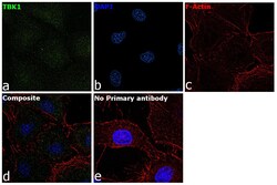
- Immunofluorescence analysis of TBK1 was performed using 70% confluent log phase A549 cells. The cells were fixed with 4% paraformaldehyde for 10 minutes, permeabilized with 0.1% Triton™ X-100 for 15 minutes, and blocked with 2% BSA for 1 hour at room temperature. The cells were labeled with TBK1 Polyclonal Antibody (Product # PA5-17478) at 1:100 dilution in 0.1% BSA, incubated at 4 degree celsius overnight and then labeled with Goat anti-Rabbit IgG (H+L) Superclonal™ Recombinant Secondary Antibody, Alexa Fluor® 488 (Product # PA5-17478) at a dilution of 1:100 for 45 minutes at room temperature (Panel a: green). Nuclei (Panel b: blue) were stained with ProLong™ Diamond Antifade Mountant with DAPI (Product # P36962). F-actin (Panel c: red) was stained with Rhodamine Phalloidin (Product # R415). Panel d represents the merged image showing cytoplasmic localization. Panel e represents control cells with no primary antibody to assess background. The images were captured at 60X magnification.
Supportive validation
- Submitted by
- Invitrogen Antibodies (provider)
- Main image
- Experimental details
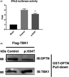
- Figure 4 In vitro functional analysis of TBK1 mutation. (a) Luciferase activity in HEK293T cells cotransfected with an IFN-beta reporter plasmid and a GFP-tagged wild-type plasmid or a p.Ile334Thr mutation TBK1 plasmid for 24 hr. Firefly luciferase activity served as an internal control. Values are expressed as mean +- SEM , ** p < 0.05. (b) Interaction between TBK1 and its downstream autophagy receptor optineurin. Lysates of a GFP-tagged wild-type TBK1 or p.Ile334Thr from HEK293T cells were incubated with GST-OPTN. Both cell lysates and bound proteins were detected by western blotting