Explore
 Validate
 Learn
Learn Western blot
Antibody data
- Antibody Data
- Antigen structure
- References [0]
- Comments [0]
- Validations
- Western blot [1]
Submit
Validation data
Reference
Comment
Report error
- Product number
- 692502 - Provider product page

- Provider
- BioLegend
- Proper citation
- BioLegend Cat#692502, RRID:AB_2632765
- Product name
- Purified anti-IL-12/IL-23 p40
- Antibody type
- Monoclonal
- Description
- IL-12 and IL-23 share the p40 subunit, which heterodimerizes respectively with IL-12 p35 or IL-23 p19 subunits to form IL-12 or IL-23. IL-12 p40 exists as a monomer and as a homodimer (IL-12 p80). IL-12 acts as a growth factor for activated human T and NK cells, enhances the lytic activity of human NK cells, and stimulates the production of IFN-γ by resting human PBMC. IL-12R is formed by two chains, IL-12Rβ1 and IL-12Rβ2. IL-12Rβ1 is associated with the Janus kinase (Jak) Tyk2 and binds IL-12 p40; IL-12Rβ2 is associated with Jak2 and binds either the heterodimer or the p35 chain. Signaling through the IL-12 receptor complex induces phosphorylation, dimerization, and nuclear translocation of several signal transducers and activators of transcription (STAT) family members (STAT1, 3, 4, 5), but most of the biological responses to IL-12 have been attributed to STAT4. IL-12 has been shown to elicit anti-tumor activity in mice and humans. It is believed that the antitumor effects of IL-12 are mediated, at least in part, by indirect mechanisms. Induction of IFN-γ results in the upregulation of class I and class II MHC molecules, adhesion molecules (ICAM-1), nitric oxide production by antigen presenting cells (APC), and the production of additional cytokines, CXCL9 and 10, which in turn mediate angiostatic effects.
- Reactivity
- Human
- Host
- Mouse
- Conjugate
- Unconjugated
- Antibody clone number
- A15076D
- Vial size
- 100 µg
- Concentration
- 0.5 mg/ml
- Storage
- 2°C-8°C
- Handling
- Ambient RT
No comments: Submit comment
Supportive validation
- Submitted by
- BioLegend (provider)
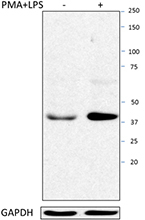
- Main image
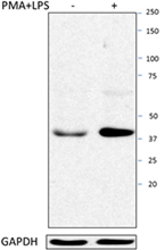
- Experimental details
- THP-1 cells were non-treated or treated with 100 ng/ml PMA for 3 days, and then treated with 1 µg/ml LPS for 1 day. Total protein extracts (15 µg protein) were resolved by 4-12% Bis-tris gel electrophoresis, transferred to nitrocellulose, and probed with 1 µg/mL purified anti-IL-12/IL-23 p40 (clone A15076D) antibody. Proteins were visualized using a goat anti-mouse-IgG secondary antibody conjugated to HRP or donkey anti-rabbit IgG secondary antibody conjugated to HRP and chemiluminescence detection. Purified anti-GAPDH (clone Poly6314) antibody was used as a loading control.
- Conjugate
- Unconjugated