Explore
 Validate
 Learn
LearnHPA001040
antibody from Atlas Antibodies
Targeting: FHL1
bA535K18.1, FHL1B, FLH1A, KYO-T, MGC111107, SLIM1, XMPMA
 Western blot
 Immunocytochemistry
Antibody data
- Antibody Data
- Antigen structure
- References [2]
- Comments [0]
- Validations
- Immunocytochemistry [1]
- Immunohistochemistry [1]
Submit
Validation data
Reference
Comment
Report error
- Product number
- HPA001040 - Provider product page

- Provider
- Atlas Antibodies
- Proper citation
- Atlas Antibodies Cat#HPA001040, RRID:AB_1848541
- Product name
- Anti-FHL1
- Antibody type
- Polyclonal
- Description
- Polyclonal Antibody against Human FHL1, Gene description: four and a half LIM domains 1, Alternative Gene Names: bA535K18.1, FHL1B, FLH1A, KYO-T, MGC111107, SLIM1, XMPMA, Validated applications: WB, IHC, ICC, Uniprot ID: Q13642, Storage: Store at +4°C for short term storage. Long time storage is recommended at -20°C.
- Reactivity
- Human
- Host
- Rabbit
- Conjugate
- Unconjugated
- Isotype
- IgG
- Vial size
- 100 µl
- Concentration
- 0.1 mg/ml
- Storage
- Store at +4°C for short term storage. Long time storage is recommended at -20°C.
- Handling
- The antibody solution should be gently mixed before use.
Submitted references Molecular autopsy and family screening in a young case of sudden cardiac death reveals an unusually severe case of FHL1 related hypertrophic cardiomyopathy
From Gene Expression Analysis to Tissue Microarrays
Gaertner‐Rommel A, Tiesmeier J, Jakob T, Strickmann B, Veit G, Bachmann‐Mennenga B, Paluszkiewicz L, Klingel K, Schulz U, Laser K, Karger B, Pfeiffer H, Milting H
Molecular Genetics & Genomic Medicine 2019;7(8)
Molecular Genetics & Genomic Medicine 2019;7(8)
From Gene Expression Analysis to Tissue Microarrays
Ek S, Andréasson U, Hober S, Kampf C, Pontén F, Uhlén M, Merz H, Borrebaeck C
Molecular & Cellular Proteomics 2006;5(6):1072-1081
Molecular & Cellular Proteomics 2006;5(6):1072-1081
No comments: Submit comment
Supportive validation
- Submitted by
- Atlas Antibodies (provider)
- Main image

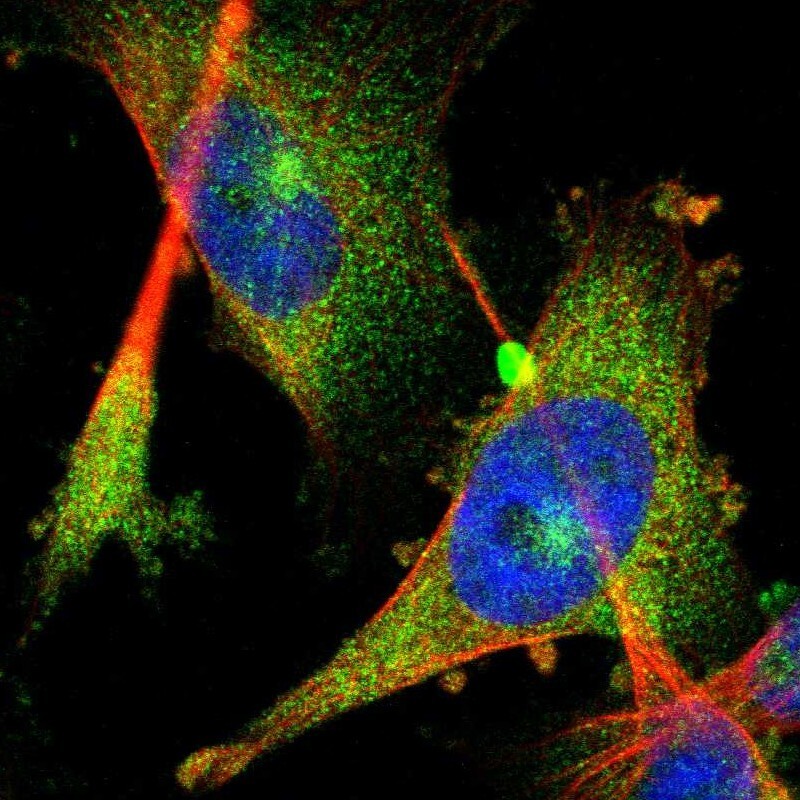
- Experimental details
- Immunofluorescent staining of human cell line U-251 MG shows localization to cytosol.
- Sample type
- Human
Supportive validation
- Submitted by
- Atlas Antibodies (provider)
- Enhanced method
- Orthogonal validation
- Main image
- Experimental details
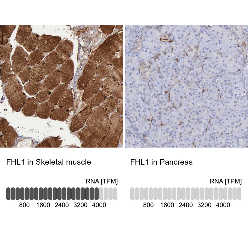
- Immunohistochemistry analysis in human skeletal muscle and pancreas tissues using HPA001040 antibody. Corresponding FHL1 RNA-seq data are presented for the same tissues.
- Sample type
- Human
- Protocol
- Protocol