Explore
 Validate
 Learn
LearnHPA045827
antibody from Atlas Antibodies
Targeting: CD40LG
CD154, CD40L, gp39, hCD40L, HIGM1, IMD3, TNFSF5, TRAP
 Western blot
 Immunohistochemistry
Antibody data
- Antibody Data
- Antigen structure
- References [1]
- Comments [0]
- Validations
- Western blot [1]
- Immunohistochemistry [1]
Submit
Validation data
Reference
Comment
Report error
- Product number
- HPA045827 - Provider product page

- Provider
- Atlas Antibodies
- Proper citation
- Atlas Antibodies Cat#HPA045827, RRID:AB_10959606
- Product name
- Anti-CD40LG
- Antibody type
- Polyclonal
- Description
- Polyclonal Antibody against Human CD40LG, Gene description: CD40 ligand, Alternative Gene Names: CD154, CD40L, gp39, hCD40L, HIGM1, IMD3, TNFSF5, TRAP, Validated applications: WB, IHC, Uniprot ID: P29965, Storage: Store at +4°C for short term storage. Long time storage is recommended at -20°C.
- Reactivity
- Human
- Host
- Rabbit
- Conjugate
- Unconjugated
- Isotype
- IgG
- Vial size
- 100 µl
- Concentration
- 0.1 mg/ml
- Storage
- Store at +4°C for short term storage. Long time storage is recommended at -20°C.
- Handling
- The antibody solution should be gently mixed before use.
Submitted references Incomplete Freund’s adjuvant reduces arginase and enhances Th1 dominance, TLR signaling and CD40 ligand expression in the vaccine site microenvironment
Pollack K, Meneveau M, Melssen M, Lynch K, Koeppel A, Young S, Turner S, Kumar P, Sol-Church K, Mauldin I, Slingluff Jr C
Journal for ImmunoTherapy of Cancer 2020;8(1):e000544
Journal for ImmunoTherapy of Cancer 2020;8(1):e000544
No comments: Submit comment
Enhanced validation
- Submitted by
- Atlas Antibodies (provider)
- Enhanced method
- Recombinant expression validation
- Main image
- Experimental details
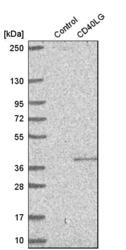
- Western blot analysis in control (vector only transfected HEK293T lysate) and cD40LG over-expression lysate (Co-expressed with a C-terminal myc-DDK tag (~3.1 kDa) in mammalian HEK293T cells, LY400020).
- Sample type
- Human
- Protocol
- Protocol
Supportive validation
- Submitted by
- Atlas Antibodies (provider)
- Enhanced method
- Orthogonal validation
- Main image
- Experimental details
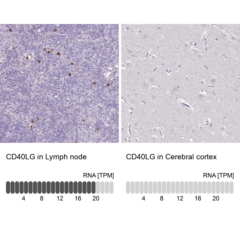
- Immunohistochemistry analysis in human lymph node and cerebral cortex tissues using HPA045827 antibody. Corresponding CD40LG RNA-seq data are presented for the same tissues.
- Sample type
- Human
- Protocol
- Protocol