Explore
 Validate
 Learn
Learn14-1548-82
antibody from Invitrogen Antibodies
Targeting: CD40LG
CD154, CD40L, gp39, hCD40L, HIGM1, IMD3, TNFSF5, TRAP
 Flow cytometry
 Other assay
Antibody data
- Antibody Data
- Antigen structure
- References [6]
- Comments [0]
- Validations
- Other assay [3]
Submit
Validation data
Reference
Comment
Report error
- Product number
- 14-1548-82 - Provider product page

- Provider
- Invitrogen Antibodies
- Product name
- CD154 (CD40 Ligand) Monoclonal Antibody (24-31), eBioscience™
- Antibody type
- Monoclonal
- Antigen
- Other
- Description
- Description: The 24-31 monoclonal antibody reacts with human CD154, a 39 kDa transmembrane glycoprotein also known as gp39 and CD40 ligand (CD40L). CD154 is a member of the TNF superfamily and is expressed transiently by activated T cells. Through its binding to CD40 on antigen presenting cells including B cells, monocytes/macrophages and dendritic cells, CD154 serves a crucial function in T-APC cognate interaction. CD154 interaction with CD40 transduces signals for T-dependent B-cell activation and induces B cell cycle entry. 24-31 cross-reacts with the cynomolgus monkey CD154. Applications Reported: 24-31 has been reported for use in flow cytometric analysis. 24-31 has also been reported to block CD154 binding to CD40 and inhibit T cell-dependent B cell differentiation and antibody response in vitro. (Please use Functional Grade Purified 24-31, Product # 16-1548, in functional assays). Applications Tested: The 24-31 antibody has been tested by flow cytometric analysis of resting and activated human normal human peripheral blood cells. This can be used at less than or equal to 1 µg per test. A test is defined as the amount (µg) of antibody that will stain a cell sample in a final volume of 100 µL. Cell number should be determined empirically but can range from 10^5 to 10^8 cells/test. It is recommended that the antibody be carefully titrated for optimal performance in the assay of interest. Purity: Greater than 90%, as determined by SDS-PAGE. Aggregation: Less than 10%, as determined by HPLC. Filtration: 0.2 µm post-manufacturing filtered.
- Reactivity
- Human
- Host
- Mouse
- Isotype
- IgG
- Antibody clone number
- 24-31
- Vial size
- 100 µg
- Concentration
- 0.5 mg/mL
- Storage
- 4° C
Submitted references Functional SARS-CoV-2-Specific Immune Memory Persists after Mild COVID-19.
Photodynamic therapy reduces the inhibitory effect of osteosarcoma cells on dendritic cells by upregulating HSP70.
Optimization of mass cytometry sample cryopreservation after staining.
CD4-like immunological function by CD4- T cells in multiple natural hosts of simian immunodeficiency virus.
Comparative analysis of MVA-CD40L and MVA-TRICOM vectors for enhancing the immunogenicity of chronic lymphocytic leukemia (CLL) cells.
A humanized anti-human CD154 monoclonal antibody blocks CD154-CD40 mediated human B cell activation.
Rodda LB, Netland J, Shehata L, Pruner KB, Morawski PA, Thouvenel CD, Takehara KK, Eggenberger J, Hemann EA, Waterman HR, Fahning ML, Chen Y, Hale M, Rathe J, Stokes C, Wrenn S, Fiala B, Carter L, Hamerman JA, King NP, Gale M Jr, Campbell DJ, Rawlings DJ, Pepper M
Cell 2021 Jan 7;184(1):169-183.e17
Cell 2021 Jan 7;184(1):169-183.e17
Photodynamic therapy reduces the inhibitory effect of osteosarcoma cells on dendritic cells by upregulating HSP70.
Zhang F, Zhu Y, Fan G, Hu S
Oncology letters 2018 Oct;16(4):5034-5040
Oncology letters 2018 Oct;16(4):5034-5040
Optimization of mass cytometry sample cryopreservation after staining.
Sumatoh HR, Teng KW, Cheng Y, Newell EW
Cytometry. Part A : the journal of the International Society for Analytical Cytology 2017 Jan;91(1):48-61
Cytometry. Part A : the journal of the International Society for Analytical Cytology 2017 Jan;91(1):48-61
CD4-like immunological function by CD4- T cells in multiple natural hosts of simian immunodeficiency virus.
Vinton C, Klatt NR, Harris LD, Briant JA, Sanders-Beer BE, Herbert R, Woodward R, Silvestri G, Pandrea I, Apetrei C, Hirsch VM, Brenchley JM
Journal of virology 2011 Sep;85(17):8702-8
Journal of virology 2011 Sep;85(17):8702-8
Comparative analysis of MVA-CD40L and MVA-TRICOM vectors for enhancing the immunogenicity of chronic lymphocytic leukemia (CLL) cells.
Litzinger MT, Foon KA, Tsang KY, Schlom J, Palena C
Leukemia research 2010 Oct;34(10):1351-7
Leukemia research 2010 Oct;34(10):1351-7
A humanized anti-human CD154 monoclonal antibody blocks CD154-CD40 mediated human B cell activation.
Brams P, Black A, Padlan EA, Hariharan K, Leonard J, Chambers-Slater K, Noelle RJ, Newman R
International immunopharmacology 2001 Feb;1(2):277-94
International immunopharmacology 2001 Feb;1(2):277-94
No comments: Submit comment
Supportive validation
- Submitted by
- Invitrogen Antibodies (provider)
- Main image
- Experimental details
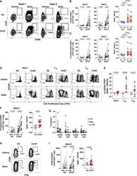
- Figure 3 SARS-CoV-2 Infection Induces Durable, Functional Spike-Reactive CD4 + T Cells (A) Representative flow cytometry plots of ICOS and CD40L expression on antigen-experienced (non-CD45RA + CCR7 + ) CD4 + T cells 20 h after incubation of PBMCs from HC and CoV2 + individuals at V1 and V2 with vehicle or SARS-CoV-2 spike. (B) Number of antigen-experienced ICOS + CD40L + CD4 + T cells per 1x10 6 CD4 + T cells from HC and CoV2 + samples after incubation with vehicle (Veh.) or spike (S) at both time points (right) and calculated number of spike-responsive CD4 + T cells (number after incubation with spike minus number after incubation with vehicle) compared across time points (left). (C) Number of antigen-experienced CXCR5 + ICOS + CD40L + CD4 + T cells (cTfh) per 1 x 10 6 CD4 + T cells from HC and CoV2 + samples after incubation with Veh. or S at both time points (right) and calculated number of spike-responsive cTfh cells (number after incubation with spike minus number after incubation with vehicle) compared across time points (left). (D) Representative flow cytometry plots of sorted CD4 + naive (CD45RA + CCR7 + ), T CM (CD45RA - CCR7 + ), or T EM (CD45RA - CCR7 - ) T cells from HC and CoV2 + PBMCs after 5-6 days of co-culture with SARS-CoV-2 spike-protein-pulsed autologous monocytes and measuring proliferation by CPD dilution. (E) SARS-CoV-2 spike-specific expansion of sorted CD4 + naive T, T CM , and T EM cells from V1 (circles) and V2 (squares) reported as frequency of CXC
- Submitted by
- Invitrogen Antibodies (provider)
- Main image
- Experimental details
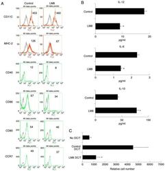
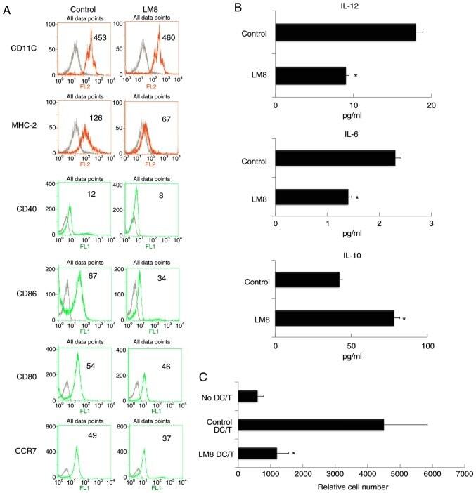
- Figure 1. The remnants of LM8 decreased the co-stimulatory molecules, and inhibited IL-12 and IL-6 levels, increased IL-10 levels, and inhibited the ability of DCs to stimulate T-cell proliferation. (A) The DCs were isolated following a 48-h co-culture with the remnants of LM8 cells. The cells were then labeled with antibodies against CD11c, MHC-2, CD40, CD86, CD80 and CCR7 for phenotypic analysis by flow cytometry. The numbers in the histograms indicate the geometric mean fluorescence intensity. (B) Following isolation from the co-culture system, the DCs were cultured for 12 h. Subsequently, the expression levels of IL-12, IL-6 and IL-10 in the supernatant were analyzed by ELISA. (C) CD4 + T cells from DO11.10 OVA 323-339 -specific (TCR-transgenic x C57BL/6) F1 hybrid mice were co-cultured with DCs or mDCs (control) in the presence of OVA peptides. Five days later, the total number of viable CD4 + T (CD4 + 7-AAD - ) cells in each well was measured by flow cytometry. Data represent one of at least three experiments with similar results. *P
- Submitted by
- Invitrogen Antibodies (provider)
- Main image
- Experimental details
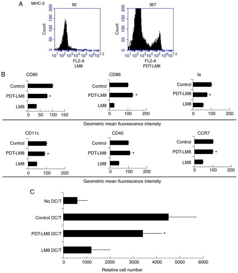
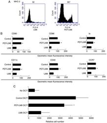
- Figure 2. PDT treatment partly reversed the effect of LM8 remnants on the phenotype of DCs and their ability to stimulate T cells proliferation. (A) The LM8 cells were pre-treated with PDT, and then labeled with antibodies against MHC-2, CD11c, CD40, CD86, CD80 and CCR7, for phenotypic analysis by flow cytometry. (B) The numbers in the histograms indicate the geometric mean fluorescence intensity. *P